INTRODUCTIONIn otorhinoloryngology, there are many different types of acute and chronic infections which are responsible for conditions more severe or not because of factors such as virulence of infectious agent, its particle number and host condition. Among these infections, rhinosinusites are the most frequent and severe ones, which remain with high rates of morbiness and mortality, still prevailing on poor population, but with considerable incidence even in developed countries and with godd level of health assitance (1).
Rhinosinusitis can be described as inflamation of rhinosinusal mucosa in response to action of infectious, traumatic and chemical events or even action of substances that can cause allergic reaction, causing an inflamatory state of mucosa. Such event has acute features which can be solved spontaneously or by medication action that will interact in order to adjust nasal mucosa and paranasal sinus. Therefore, in some cases, this does not occur and persistance of these alterations leads to a chronic state (2).
The coming of different efficacious antimicrobials against etiological agents of nasosinusal infections expressively contributed to reduce incidence of their complications. Therefore, because of wide and improper use of such medication, it has been emerging resistent bacterias, having as consequences the appearing of complicated cases of these diseases. Besides, we can mention the increasing of immune depressed patients, such as AIDS and patients who underwent any kind of transplantation (3).
Among intracranial infectious complications of nasosinusal origin, there are the ones which take spaces: intracranial abscess (ICA), subdural empyema (SDE) and epidural abscess (EDA). The purpose of the current study was to retrospectively analyze intracranial infectious complications resulted from rhinosinusitis, in hospitalized patients at Serviço de Neurocirurgia do Hospital Governador João Alves Filho-HGJAF, Aracaju-SE (Service of Neurosurgery of Governador João Alves Hospital), in the period of January 1995 to September 2004.
RECORDS AND METHODSIt was retrospectively analyzed 21 patients, males and females, with secondary intracranial complications and nasosinusal infectious, assisted at Serviço de Neurocirurgia do Hospital Governador João Alves Filho-HGJAF, Aracaju-SE, in the period of January 1995 to September 2004.
In the mentioned study patients who had their records with evidences of nasosinusal infectious with intracranial complication, confirmed through computed tomography scan and/or magnetic resonance imaging were included. Patients were analyzed according to sex, age, clinical condition, type of intracranial complication, neuroimaging exams, treatment, prognosis and sequelae. All the information was registered in separeted forms. The study had the approval from Ethics Committee in Research of Hospital Universitário da Universidade Federal de Sergipe, protocol number 105/04.
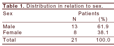
It is clearly noticed that in a total of 21 patients who presented intracaranial complications due to rhinosinusitis, 13 of them (61.9%) were male and eight (38.1%) were female (Table1).
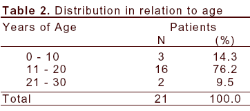
They aged between 5 and 29, and the average was 15.8 years of age. There were 16 patients (76.2%) aging between 11 and 20, three (14.3%) upt to ten years, and two (9.5%) between 21 and 30 years (Table 2).
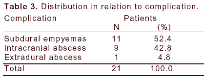
In relation to frequency of complications, there is SDE with frequency of 52.4% from studied patients, while ICA was diagnosed in 42.8% of the cases and one patient (4.8%) presented EDA (Table 3).
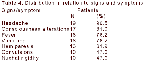
The symptoms presented were an association of intracranianal hypertesion syndromes and infectious syndrome. The most common symptom was headache in 19 cases (90.5%), followed by alteration on conscious level in 17 cases (81%). Fever and vomiting were present in 16 (76.2%), hemiparesia in 13 (61.9%), convulsion and nuchal rigidity in 10 cases (47.6%) (Table 4).
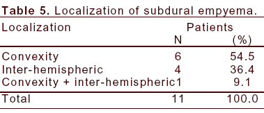
In relation to topographic distribution of lesions, from 11 cases of SDE, six of them on cerebral convexity, four interhemispheres and in one case there was association of the two localizations (Table 5)
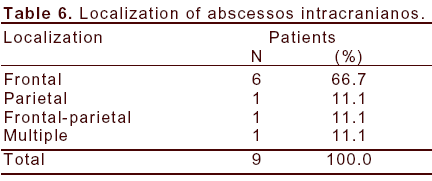
In relation to ICA, from 9 cases, six of them were placed on frontal lobe, one case on parietal lobe, one case on frontal-parietal area, and one case of multiple ICA (Table 6). The only case of EDA in our series was placed on frontal area.
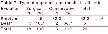
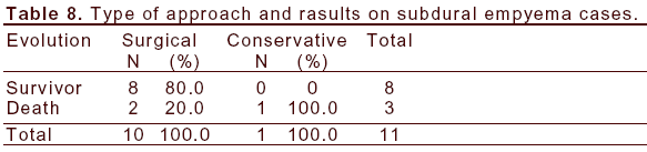
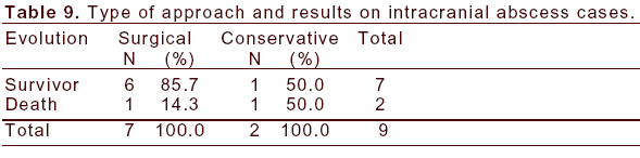
The surgical approach was established in 18 patients (85.7%) while the other three patients (14.3%) were treated clinically. In surgical group, mortality reached 16.7%, while in the group treated in a conservative manner, such rate reached 66.7% (Table 7). Among the 11 cases of SDE, 10 were submitted to surgical treatment, and eight of them (80%) survived. Only one patient was treated in a conservative manner, but died (Table 8). In relation to the nine cases of ICA, seven patients were submitted to surgery, having 85.7% of survival, while from the two patients treated clinically, one (50%) died (Table 9). The patient with EDA was submitted to surgical treatment and survived.
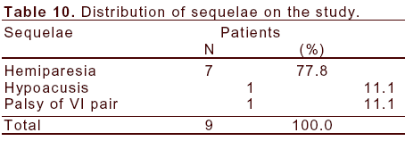
When being released from hospital, seven patients presented no sequelae, but nine of them did. The sequelae were: hemiparesia (7 cases), hypoacusis (1 case) and palsy of the sixth cranial pair (1 case) (Table 10).
DISCUSSIONIn our study, there was a predominance of 13 male cases and 8 female cases, what agreess with may authors (4-8), without any explantions for that (9,10).
The distribution of the patients by age showed greater incidence in the second decade of life. This information was found in many studies (6,9,11,12). According to KERR et al. (1958) (13), this fact can be assigned to greater frequencies of infections of paranasal sinus in the second decade of life. KAPLAN (1976) (14) suggested that intracranial complications during adolescence occurs due to larger bloody flow on diploic avalvular system and by continous growth of frontal sinus, making its posterior wall a fine barrier to nasosinusal infections. Therefoe, other authors found a high incidence in older patients (4,5,7).
Among intracranianal complications studied in this work, we can observe predominance of SDE (52.4%), followed by ICA (42.8%) and EDA (4.8%). The medical literature diverges as for intracranianal complications more frequent in the course of rhinosinusites. To some authors is the ICA (9,11), for others is the EDA (5) or SDE (6.12). However, rhinosinusites are mentioned in the literature as the main cause of SDE (15-17).
In our work, we did not study the presence of causal microorganism, though the records cover hospitalized patients since 1995, when culture of secretions was not done in routine way. Besides, even today, we have difficulties to the accomplishment of culture to anaerobic microorganism. Among cultures done on cases of the current study, there was no growth of microorganism, perhaps by previous and extended use of antibiotics, fact that is also observed by other authors (5,9,18).
The most frequent symptom in our study was headache, followed by alterations on consciousness level, fever and vomitting. Hemiparesia, convulsions and nuchal rigidity were less frequent. As reported in the literature, the symptom of intracranial complications coming from rhinosinusitis is usually an association of infectious syndrome, intracranial hypertension syndrome and focal neurological alterations, which can express themselves simultaneously, with superposition of their signs and symptoms (3-5,7-9,11,19-21). The literature does not agree on those most frequent symptoms, nevertheless different authors mention headache and fever as most common clinical manifestations, though they are unspecified. So patients should be evaluated as for nuchal rigidity, presence of focal neurological signs and alterations on consciousness level.
In relation to localization of SDE, six (54.5%) are on convexity, followed by inter-hemispheres, with four cases (36.4%), what was also reported by other authors (10,15,19,22,23). According to FONSECA AND RODRIGUES (1993) (15), this occurs because subdural spaces is free on these areas and restricted on cranio basis.
In relation to localization of ICA, 66.7% of our cases were presented at frontal lobe, what agrees with the literature, which states this localization as preferential, no matter what paranasal cavities are implicated.
In relation to EDA, C.T. showed subdural hypodensity on interhemispheric fissure associated to frontal epidural hypodensity, while M.R. with gadolinium showed EDA placed on convexity, through cerebral sulcus, what shows the reliability of this exam on lesions diagnosis, as demonstrated by other authors.
In relation to the established treatment, three patients were treated in conservative manner, one of the cases, due to reduced amount of pus, and the other two due to uncertain general conditon of patient. Those two died, testifying 66.7% of mortality rate in this group. As testified by other authors, few colections can be clinically treated with satisfatory results (3,4,24). In 18 patients was applied surgical procedure, and punction through trepanation was established in nine cases and craniotomy with drainage of lesion on the nine ones remaining. Three of them (16.7%) in this group died. Among death cases, two of them were submitted to punction through trepanation and one to craniotomy. As reported in other studies, there was no big difference on the prognosis among patients submitted to one of these surgical techniques (25). When analyzing separetely the intracranial complications, we notice that two or three patients submitted to conservative treatment presented ICA. Among those, one was not submitted to surgery due to bad clinical conditions, diying afterwards. Only one case of SDE was treated in a conservative manner, due to bad genaral condition, and patient also died. The patient with EDA presented good clinical conditions, and was submitted to surgery successfully. In this way, we may understand that it was not lack of surgical treatment that influenced high mortality in the group treated in a conservative manner, but bad general conditions at the moment of hospitalization, what was reported by BORRÁS et al.(2002) (4). The high number of patients in bad clinical conditions in our records can be explained by the fact that HGJAF (hospital) assist large part of poor people from different states of North of Brazil (Sergipe, Alagoas, Bahia and Pernanbuco), and it lacks level of health assitance, and when patients are sent to our service is sometimes late.
Among the patients who survived, nine (56.25%) presented sequelae. This rate is considered high when compared to results of other authors (3,4,7,21). Therefore, in these works was done a follow-up of such patients, what might have contributed to a gradual recovery of the sequelae, while in the current study, it was not possible to follow patients up in the ambulatory, though we evaluated sequelae when in high level. Among them, hemiparesia was the most frequent, occurring on seven cases, what has been already showed in other works (9,16). To other authors (3,6), convulsion crises presented more incidence. Other sequelae observed in our series, were hypoacusis and facial palsy, both in one case.
CONCLUSION1 - Intracranial complications coming from rhinosinusitis which presented higher incidence was SDE (52.4%), followed by AIC (42.8%) and by EDA (4.8%).
2 - Infectious intracranial complications coming from nasosinusal were more frequent on men (61.9%).
3 - Second decade of life was more affected (76.2%).
4 - ICA was present on frontal lobe in six cases (66.7%).
5 - SDE was present with more frequency on cerebral convexity (54.5%).
6 - The most common symptom was headache (90.5%), followed by alteration on consciousness level (81%), fever (76.2%) and vomitting (76.2%).
7- Patients submitted to surgical treatment presented better prognosis, not counting type of complication.
8 - Among sequelea, hemiparesia was more frequent (77.8 %), followed by hypoacusis (11.1%) and palsy of VI cranial pair (11.1%).
BIBLIOGRAPHY1. Lopes Filho O, Campos CAH. Tratado de Otorrinolarimgologia. 1ª ed. São Paulo: Roca; 1994.
2. Dorgan JV, Souza BB, Sarreta SMC, Ferreira MDS, Melo VR, Lima WTA. Estudo histológico e ultraestrutural da mucosa do seio maxilar em pacientes com rinossinusite crônica e polipose nasossinusal. Rev Bras Otorrinilaringol, 2004, 70: 7-13.
3. Roche M, Humphreys H, Smyth E, Cunney R, McNara E, O'Brien D et al. A twelve-year review of central nervous system bacterial abscesses; presentation and aetiology. Clin Microbiol Infect, 2003, 9: 803-9.
4. Borrás JM, Garcia-Bach M, Maestro-de León JI, Aparicio A, Juan N, Martínez-Lacasa J et al. Coleciones purulentas intracraneales. Revision de 34 casos tratados quirúrgicamente a lo longo de doce años (1989-2000). Neurocirurgía, 2002, 1: 6-14.
5. Gallagher RM, Gross CW, Phillips CD. Suppurative intracranial complications of sinusitis. Laryngoscope, 1998, 108: 1635-1642.
6. Jones NS, Walker JL, Bassi S, Jones T, Punt J. The intracranial complications of rhinosinusitis: can they be prevented? Laryngoscope, 2002, 112: 59-63.
7. Lu CH, Chang WN, Lin YC, Tsai NW, Liliang PC, Su TM et al. Bacterial brain abscess: microbiological features, epidemiological trends and therapeutic outcomes. Q J Med, 2002, 95: 501-09.
8. Tattevin P, Bruneel F, Clair B, Lellouche F, Broucker T, Chevret S et al. Bacterial brain abscesses: a retrospective study of 94 patients admitted to an intensive care unit (1980-1999). Am J Med, 2003, 111: 143-146.
9. Clayman GL, Adams GL, Paugh DR, Koopman CF. Intracranial complications of paranasal sinusites: a combined institucional review. Laryngoscope, 1991, 101: 234-239.
10. Nathoo N, Nadvi SS, Vandellen JT, Gouws E. Intracranial subdural empiema in the era of computed tomography: a review of 699 cases. Neurosurgery, 1999, 44: 529-35.
11. Giannoni C, Sulek M, Friedman EM. Intracranial complications of sinusitis: a pediatric series. A J Rhinol, 1998, 12: 173-8.
12. Singh B, Van Dellen J, Ramjettan S, Maharaj TJ. Sinogenic intracranial complications. J Laryngol Otol, 1995, 109: 945-50.
13. Kerr FWL, King RB, Meagher JN. Brain abscess: a study of 47 consecutive cases. JAMA, 1958, 168: 868-72.
14. Kaplan RJ. Neurological complications of infections of the head and neck. Otolaryngol Clin North Am, 1976, 9: 729-49.
15. Fonseca ALV, Rodrigues FF. Empiema subdural. Rev Bras Neurol, 1993, 29: 17-22.
16. Hoyt DJ, Fisher SR. Otolaryngologic management of patients with subdural empyema. Laryngoscope, 1991, 101: 20-4.
17. Wackym PA, Canalis RF, Feuerman T. Subdural empyema of otorhinological origin. J Laryngol Otol,1990, 104: 118-122.
18. Kangsanarak J, Navacharoen N, Fooanant S, Ruckphaopunt K. Intracranial complications of suppurative otitis media: 13 years experience. Am J Otolaryngol, 1995, 16: 104-9.
19. Dill SR, Coobs CG, McDonald CK. Subdural empyema: analysis of 32 cases and review. Clin Infect Dis, 1995, 20: 372-86.
20. Su TM, Lam CH, Tsai YD, Lee TC, Lu CH, Chang WN. Multiloculated pyogenic brain abscess: experience in 25 pacients. Neurosurgery, 2003, 52: 1075-80.
21. Yougev R, Bar-Meir M. Management of brian abscesses in children. Pediatr Infect Dis J, 2004, 23: 157-60.
22. Pathak A, Sharma BS, Mathuriya SN, Khosla VK, Khandelwal N, Kak VK. Controversies in the management of subdural empyema: a study of 41 cases with review of literature. Acta Nuerochir (Wien), 1990, 102: 25-32.
23. Bok APL, Peter JC. Subdural empyema: burr holes or craniotomy? J Neurosurg, 1993, 78: 574-8.
24. Garcia AB, Vázquez EG, Benito N, Górgolas M, Muñiz J, Gadea I et al. Abscesso cerebral. Estudio clinico¬mi¬cro-¬biológico y análisis pronóstico de 59 casos. Rev Clín Esp, 1998, 198: 413-9.
25. Magliani VQ, Lucantoni D, D'arrigo C, Galzio RJ. Cerebral abscesses: topic treatment with antibiotics. J Neurosurg Sci, 1988, 32: 47-50.