INTRODUCTIONFirearm injuries have been increasing worldwide (1). And Brazil, unfortunately, is not out of this context, on the contrary, more than 32,000 deaths caused by this type of accident is estimated in a year.
Injuries in the head and neck are not rare and has high rate of morbidity and mortality. The incidence on the nasal pyramid is not rare either, therefore, firearm projectile report as nasal foreign body is hardly found in the literature (5,6).
Dayal and Singh (6) reported a case of a patient who had an accident by firearm. The patient had her face seriously injured, and it was reported, through radiology, the presence of a metal object in the rhinopharinx. On the other hand, spontaneous migration from foreign bodies, either metallic or not, through tissues from the organism is described and nasal fossa can be part of their course or even their final stop.
It is reported, in this study, a case of a 30-year-patient with history of firearm projectile accident in right occipital region 2 years ago, with evolution of the presence of the projectile in the interior of right inferior nasal concha.
CASE REPORTA 30-year-male patient (from Salvador BA), searched the ENT ambulatory at the Hospital Universitário São Francisco complaining of bilateral nasal obstruction, with a predominance to the right side, associated with pain over the right maxillary region , and no other complaints. Symptoms became worse two years ago, after a firearm accident. According to patient there was only one opening in the occipital area. Patient also reported the projectile was not removed from it.
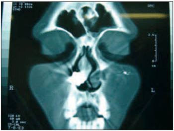
After ENT examination it was observed, at nasofibroscopy hypertrophy, septum deviation grade 3, area 2 high at right and grade 2, area 2 low at left, and also inferior nasal conchas the right. CT from paranasal sinus was required, and it showed the presence of a metal-like hyperdense image in the right nasal fossa, in the topography of inferior concha, and not presenting any images of fragments in the other areas (Picture 1).

Picture 1. CT from paranasal sinus in coronal cut.
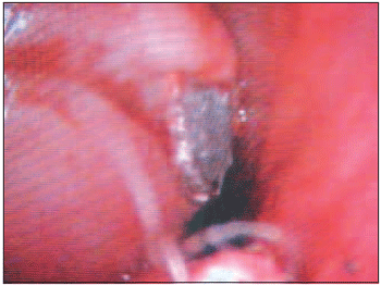
After surgery being recommended, bilateral septoplasty and removal of the projectile from the interior of the right inferior nasal concha by endoscopy were performed. It was done superficial incision in the mucosa from the superior part of concha, just to visualize the projectile. This was easily removed by a simple tension (Picture 2). Eventually, bilateral partial inferior turbinectomy, which was also performed through endoscopy, ended the procedure. Nose packing was not necessary. During anesthesia, 2gr of Cefazoline (EV) was prepared and patient made use of Cefalexin (2gr/day VO) in the first 7 days after surgery. The projectile exam showed the firearm was a type 38 rifle. The condition of the patient after surgery was satisfactory.

Picture 2. Metal projectile after incision on superior part of right inferior nasal concha.
Nasal obstruction carries different causes such as septum deviation and nasal concha hypertrophy. Although less common, the presence of a foreign body in the interior of nasal fossa can also be one of the causes (9,10,11).
Despite the fact that the incidents by firearms have been in high numbers nowadays, few cases present projectile as a foreign bodies in the nasal fossa.
In the literature by Dayal and Singh (6), only one case reported the presence of a metal object in the rhinopharynx.
The diagnosis from nasal foreign bodies is simple. Besides unilateral nasal obstruction and/or hyaline or fetid purulent rhinorrhea, the ENT exam, nasofibroscopy and CT from paranasal sinus, depending on development period, reveal the diagnosis (1,10,11).
In our report, anamnesis and physical and video exams were important when revealed bilateral septum deviation and hypertrophy of the conchas to the right; therefore only after imaging exams the diagnosis was concluded. The wonder was how such projectile stored in the interior of the inferior concha with only one entrance opening in the occipital area. Or, would there be another opening? Patient assured negatively. It is amazing how it did not occur injuries on vital structures and other sequela after the accident. An acceptable explanation would be the spontaneous migration of the foreign body, which was already reported by Salvati et al (7) and by Ilkko et al (8). The absence of fragments in the CT areas is a favorable argument of the proposed explanation.
The ideal procedure in case of a foreign body presence is its immediate removal, as it is an ENT urgency (9,10). The removal of a firearm projectile in the nasal fossa is also recommended, though conservative procedure can be applied when either such object does not cause any discomfort to the patient or its removal will be more risky than benefic (1,3,4).
Our patient complained of nasal obstruction caused by bilateral septum deviation and by hypertrophy of the conchas. Such symptoms were not intense before the accident, but the presence of the foreign body intensified such complaint. In order to soothe the discomfort, we performed the removal of the projectile together with septoplasty and bilateral inferior partial turbinectomy in the same surgery. Needless to say that antibiotic therapy after surgery was essential by the risk of infection on septal cartilage.
CONCLUSIONAccidents by firearms do not cause immediate damages to patient's health, so its removal is not so essential. Such projectile can, sometimes, react as a foreign body after a possible migration through tissues. Its removal will be necessary if there are either symptoms or risk of life.
REFERENCES1. Reiss M, Reiss G, Pilling E: Gunshot injuries in the head and neck area - basic principles, diagnosis and managment. Schweiz Rundsch Med Prax, 1998, 87(24):832-8.
2. Brasil registra mais mortes por armas de fogo do que conflitos armados internacionais. Disponível em http: www.unescobrasil.org.br . Acessado em 09 de dezembro de 2005.
3. Castro C, Santos S, Fernandez R, Labella T: Non-fatal firearm injuries of the head and neck. An Otorrinolaringol Ibero Am, 1989, 16(3):257-70.
4. Ogunleye AO, Adeleye AO, Ayodele KJ, Usman MO, Shokunbi MT: Arrow injury to the skull base. West Afr J Med, 2004, 23(1):94-6.
5. Sobrinho FPG, Jardim AMB, Sant'Ana IC, Lessa HA: Corpo estranho na nasofaringe: a propósito de um caso. Rev Bras Otorrinolaringol, 2004, 70(1):24-8.
6. Dayal D, Singh AP: Foreign body nasopharynx. J Laryngol Otol, 1970, 84:1157-60.
7. Salvati M, Cervoni L, Rocchi G, Rastelli E, Delfini R: Spontaneus movement of metallic foreign bodies. Case Report. J Neurosurg Sci, 1997, 41(4):423-5.
8. Ilkko E, Reponen J, Ukkola V, Koivukangas J: Spontaneus migration of foreign bodies in the central nervous system. Clin Radiol, 1998, 53(3):221-5.
9. Hong D, Chu YF, Tong KM, Hsiao CJ: Button batteries as foreign bodies in the nasal cavities. Int J Pediatr Otorrinolaryngol, 1987, 14(1):15-9.
10. Duarte AF, Soler RC, Zavarezzi F: Endoscopia nasossinusal associada a tomografia computadorizada de seios paranasais no diagnóstico de obstrução nasal crônica. Rev Bras Otorrinolaringol, 2005, l71(3):361-3.
11. Pinheiro SD, Freitas MR. Obstrução nasal. In: Campos CAH, Costa HOO. Tratado de Otorrinolaringologia Vol 3. 1ª ed. São Paulo: Roca; 2003, p.166-74.
1. Resident Doctor at Serviço de Otorrinolaringologia do Hospital Universitário São Francisco (ENT Service)
2. PhD In Otorhinolaringology by USP (Head of the ENT Service at Hospital Universitário São Francisco)
Study done at Hospital Universitário São Francisco - Bragança Paulista.
João Alcides Miranda, Rua Bias Fortes, 438, Centro, Boa Esperança - MG, CEP 37170-000 - phone (11) 9111-5881, e-mail jamiranda78@hotmail.com.
This article was submitted to SGP - Sistema de Gestão de Publicações (Publication Management System) from RAIO on March 24, 2006 and was approved on April 24, 2006 22:30:22.