INTRODUCTION The nasal blockage is a very common complaint and it has many causes. The most common ones are: allergic rhinitis, septal deviation, nasosinusal polyps and others.
More rarely tumors can occur, which has a low incidence in children, prevailing juvenile angiofibroma and rhabdomyosarcoma.
Meningiomas are type of intracranial neoplasias, figuring from 14 to 18% of all. About 20% of intracranial meningiomas present extracranial expansion, in places such as orbit, mid ear, nasal cavity, nasopharynx and paranasal sinus(1,2,3,4).
Therefore, primary extracranial meningioma is a rare neoplasia, it is histologically identical to intracranial meningiomas. There is greater difficulty regarding its diagnosis since meningiomas of the nasosinusal area are distinguished from esthesioneuroblastomia one, squamous cell carcinoma, melanoma, hemangioma, juvenile sarcoma and angiofibroma(2,3,4,5,6).
Primary extracranial meningiomas are rare in children and occur more commonly in the women(4).
It was reported a case of a 13-year-olf girl with diagnosis of primary meningioma in nasosinusal area.
CASE REPORTA 13-year-old girl patient, searched ENT Clinic (Ambulatório de Otorrinolaringologia e Cirúrgia de Cabeça e Pescoço - Hospital of Base - FAMERP) complaing of frontal headache to the left, constant and in weight, followed by gradual proptosis in left eye and nasal blockage to the left, for 30 days. She denied fever, rhinorrhea, epistaxis and alterations on the smelling or vision acuity.
The exam presented good condition result, with moderate proptosis in left eye (Picture 1), with no signs of local phlogistic and at rinoscopy it presented mass filling the superior meatus with flat and pink color surface in left nasal cavity. Ocular motricity and vision acuity were bilateral. Nasofibrolaryngoscopy revealed that mass did not modify its volume with Valsava's maneuver.
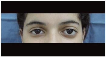
Picture 1. Picture showing proptosis in left eye.
Before this condition, patient was hospitalized and submitted to cranium and sinus of the face CT. It was observed injury with soft part aspect in the topography of the ethmoidal cells to the left determining expansion with reduction of the bone adjacent structures and no intracranial alteration was noticed (Picture 2).
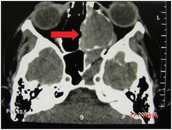
Picture 2. CT of sinus of the face: axial cut with arrow showing injury with density of soft parts in topography of ethmoidal cells to the left.
In order to plan a better surgery, it was requested a Magnetic Nuclear Resonance of Sinus of the Face and it was noticed an oval injury measuring 2 x 2cm placed in the left ethmoidal sinus, determining compressive effect on the adjacent structures with no invasion of them, with lateral deviation of the left medial rectus muscles and previous deviation of the left orbit and with intense distinction after contrast infusion (Pictures 3 and 4).
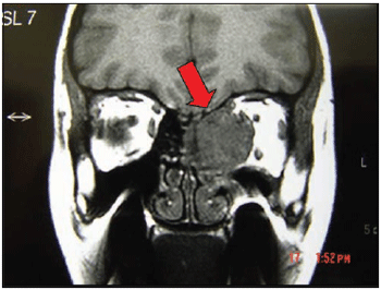
Picture 3. Nuclear Resonance Magnetic post- contrast: arrow showing oval injury in topography of ethmoidal cells to the left.
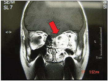
Picture 4. Nuclear Resonance Magnetic after-contrast: arrow showing oval injury in topography of ethmoidal cells to the left with intense distinction after contrast.
On 6th day in hospital, patient was submitted the surgery with combined access through External and Endonasal Ethmoidectomy. Comprehensive exeresis of the injury was performed (Picture 5). On the first postoperative day, patient presented improvement of proptosis. There were no intercurrences during or after surgery. On 8th, patient was released from hospital.
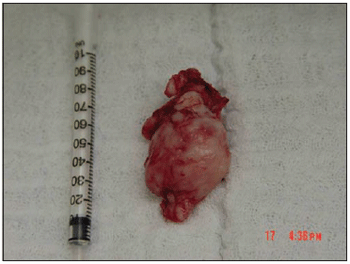
Picture 5. Picture showing injury after surgical resection.
During the clinic follow-up, it was not noticed signs of persistence of injury and histopathological exam revealed Atypical Meningioma.
DISCUSSION Primary meningioma of nasal cavity or sinus of the face is rare, with about 30 described cases in literature, and its diagnosis is difficult to be achieved. According to Petrulionis et al., tumors of epithelial origin such as squamous cell carcinoma, neurogenic origin such as esthesioneuroblastoma, odontogenic tissues such as sarcoma and ameloblastoma, vascular origin such as angiofibroma and haematological such as lymphoma must be part of the differential diagnosis(7).
Reviewing 5 cases, Friedman et al. reported that the clinical presentation depends on the localization of neoplasia, and signs and symptoms only arise after a significant growth of the injury. Complaints can be of cervical mass when located in the parapharyngeal area, mass in pre-auricular area when located in the infratemporal area, sinusitis, nasal mass, proptosis and epistaxis when located in nasal cavity or paranasal sinus(1). In our report, initial complaints were of headache frontal and proptosis, and the neoplasia was located in the etmoidal sinus.
There are some considered mechanisms to explain physiopathology of primary extracranial meningioma:
1 - Presence of arachnoid cells in nerves or vases where these emerge from the Central Nervous System.
2 - Tissue Migration of meninges for extracranial areas during embryogenesis.
3 - Traumatic event or intracranial hypertension that dislocates arachnoid cells.
4 - Origin in indifferent mesenchymal cells(1,8,9).
CONCLUSIONPrimary extracranial meningioma of nasal cavity or paranasal sinus must be part of the differential diagnosis of nasosinusal masses, even for being of rare occurrence. The diagnosis is based on anamnesis, physical exam and on complementary ones such as nasofiberlaryngoscopy, exams of image (Computerized Tomography Scan and Nuclear Magnetic Resonance) and confirmed by the anatomopathological study. The treatment for this disease is a surgical one, with good prognosis and with no need of complementary therapies(5)
REFERENCE1. Friedman CD, Constantino PD, Tietelbaum B, Berktold RE, Sisson GA. Primary extracranial meningiomas of the head and neck. Laryngoscope 1990; 100:41-8.
2. Geoffray A, Lee YY, Jing BS, Wallace S. Extracranial meningiomas of the head and neck. Am J Neuroradiol 1984; 5:599-604.
3. Taxy JB. Meningioma of paranasal sinuses. A report of two cases. Am J Surg Pathol 1990; 14:5.
4. Weinberger JM, Birt BD, Lewis AJ, Nedzelski JM. Meningioma of nasopharynx. Am J Otolaryngol 1985; 14:5.
5. Kumar S, Dringra PL, Gondal R. Ectopic meningioma of paranasal sinuses. Child Nerv Syst 1993; 9:483-4.
6. Thompson LDR, Gyure KA. Extracranial sinosal tract meningiomas. Am J Surg Pathol 2000; 24(5):640-650.
7. Petrulionis M, Valeviciene N, Paulauskiene I, Bruzaite J. Primary extracranial meningioma of the sinonasal tract. Acta Radiol 2005; 46(4):415-8.
8. Manni JJ. Ectopic meningioma of the maxillary sinus. J Laryngol Otol 1983; 97:657-60.
9. Papini M, Chiantelli A, Cantini R, et al. Nasal meningioma. Report of a case. Acta Otorhinolaryngol Belg 1989; 43:335-8.
1. Medical - 2004 (2 of years of residence in Otolaryngology and Head and Neck Surgery, - Base Hospital / FAMERP).
2. Medical 2003 (3 of years of residence in Otolaryngology and Head and Neck Surgery, - Base Hospital / FAMERP).
3. Medical 2004 (1 st year of residence in Otolaryngology and Head and Neck Surgery, - Base Hospital / FAMERP).
4. Otorhinolaryngologist Doutorando (Responsible for the Department of Surgery Craniomaxilofacial the Department of Otolaryngology and Head of Surgery
and Neck - Base Hospital / FAMERP).
5. Otorhinolaryngologist Full Professor (Head of the Department of Otolaryngology and Head and Neck Surgery - Base Hospital / FAMERP).
Hospital de Base / FAMERP (Faculty of Medicine of Sao Jose do Rio Preto) Sao Jose do Rio Preto / SP.
Mailing address: Avenida Brigadeiro Faria Lima, 5544 - Sao Jose do Rio Preto / SP - CEP 15090-000 - Phone / Fax: (17) 3201-5000 Ramal 5747 -- E-mail: danihaber@uol.com.br
This article was submitted in Management System Publications in the R@IO 3/7/2006 and approved on 1/10/2006 22:55:35