INTRODUCTIONThe carotid body is a small oval structure, with irregular surface and pinky color, measuring from 1 to 2 diameter millimeters, bilaterally located in the bifurcations of the regular carotid arteries, closely connected to the adventitia of such vessels(1).
The paraganglions are tumors which are originated from paraganglion cells of the neural crest, and develop in the paravertebral region in association with cervical blood vessels, skull nerves and autonomous nervous system(2). They may also originate from little chemoreceptor organs which are found in the adventitia of regular carotid arteries bifurcations. The paraganglions of carotid body are rare, although they make most of head and neck paraganglions.
The authors of this case taking into account such tumors' rarity, besides the surgical difficulties at exposing and resecting such lesions, which requires the participation of different medical specialties. The origin of paraganglion tumors is neurodermic and they have two kinds of cell: Cells of I kind, which contain catecolamine granules, and cells of II kind, which are similar to Schwann cells, and surround the cells of I kind. Paraganglions are highly spread into the human body, and are found in the heart, gastrointestinal tract, retroperitoneum, mediastine, lungs and bladder. In the head and neck region, its presence may be observed in the trachea, larynx, pineal gland, tongue, hypophysis and orbit. However, its most frequent localization is in the carotid body(1,3).
The objective of this case report is to present a rare case of cervical tumor emphasizing the surgical technique, the differential diagnosis and the need of multidisciplinary team participation.
LITERATURE REVIEW AND DIFFERENTIAL DIAGNOSISEach carotid body is supplied by regular and external carotid arteries branches, in a way which the chemoreceptors are always in direct contact with the arterial blood. The carotid body is highly innervated, and sympathetic nervous branches and glosso-pharyngeal nerve reach its upper portion. Small venules are also originated from that region, which end up in the tyrolingual trunk(1,2).
In 2004, Galvão et al(2) presented a case of bilateral paraganglion in the carotid body which, at the physical exam, presented a bilateral cervical mass which was hard, elastic, painless, pulsatile, vertically unmovable, horizontally movable, taking the II and III levels to the right and II level to the left. The diagnosis was confirmed by neck CT scan, Magnetic Resonance Imaging and carotid arteriography. The treatment used was surgery, in which the authors resected the lesion together with the portion of internal and external carotids which were involved. Posterior to the surgery, a reconstruction was performed with the use of Ty graft with external saphenous vein. The post-surgical phase proceeded without intercurrences, except for the paralysis of the hypoglossal nerve, which was injured once it was adhered to the tumor.
Paraganglions were primarily called chemodectomas. However, the term "carotid body tumors" arose as the most common one to design the tumors which are found in the bifurcation of carotid arteries, and are found at 0.012% of all head and neck tumors. Among the carotid body tumors, only 2% are malignant and the benignant exceptionally become malignant(4). They are originated from small chemoreceptor organs which are located in the adventitia of regular carotid arteries bifurcations. Such tumors are called paraganglions or chemodectomas(3).
França et al, in 2003, studied a group of 19 patients with a total of 20 carotid body tumors, once one of the patients presented billateral lesion. All lesions were primary and, at the physical examination, presented a pulsatile, movable and hard mass in the anterior neck triangle, with slow and progressive growing. The size of the tumors varied between 2x2 and 8x5 centimeters. The diagnosis was confirmed by neck CT scan, duplex scan and carotid arteriography. All patients were undertaken to simple surgical resection. There was no post-operatory mortality in this study group(4).
Some authors relate the hereditary feature and its bilateralism in 8% of the total(5). There is no gender prevalence and it may be diagnosed 12 and 69 years of age(5,6). If with most of the authors accepting the tumors' origin to be unknown(7), others believe that its etiology may be related to chronic hypoxia, high altitudes and dominant autosomic transmission with incomplete penetrance(6,7).
Generally, the carotid body tumors present signs which are related to its mass and with the possible compression of adjacent structures, however such local compression will not occur while the tumor is less than 3 centimeters wide(6).
Some classical signs may be observed at the physical examination, such as the finding of a hard tumor at palpation, located between the internal and external carotids (Kocher I Signal); horizontally movable tumor and vertically immovable tumor (Fontaine Sign) and, at bidigital palpation (external and intra-oral), the tumor is located in the tonsil region (Kocher II Sign)(7).
The clinical histories of patients who carry carotid body tumors are similar. The complain about the appearance of a hard, movable and pulsatile mass located in the neck anterior triangle with slow and gradual growth(8).
The carotid body tumor was first described by Taube, in 1743, who thought it was a nervous ganglion and named it "comoganglion minutun". Luschka, in 1862, Carried out the first histological description of such neoplasia and named in carotid gland(6). The first surgical resection of this tumor was carried out by Reigner, in 1880, however it ended up in obit. Maydl, in 1886, resected this tumor, but the patient became aphasic and hemiplegic. Scudder, in 1903, successfully carried out this surgery. Gordon-Taylor, in 1940, described a subadventitial disection plan which they named "white line". The greatest casuistic so far has been described by Hallet et al., in a 50-year study with 153 cases(4). The biggest tumor described was 12 centimeters wide(9).
The paraganglions differential diagnosis is carried out with lymph node enlargement, branchial cist, parotid gland tumor, thyroid tumor, neurinoma and carotid artery aneurism(10).
When no treatment is used for the carotid body tumor, its mortality may reach 30%(7). Nowadays, the elective treatment is surgery(4,9,10,11).
Radiotherapy does not offer any therapeutic aid for such lesions, and it may be used in cases of little residual post-surgical tumors(4,9). Some authors mention embolism, but they frequently do not use it. Other authors report the eventual need of artery resection and its interposition with other homologous or autologous materials(4). Surgical patients must be followed during a long period of time, once they may have metastatic diseases which may become evident in 10 or 20 years' time(3,12).
Souza et al.(9), in 2000, studied nine carotid body paraganglions in eight patients; there was one bilateral case. All patients presented a palpable mass, located below the mandible angle, which varied, from 4 up to 12 centimeters. The radiologic diagnosis was based on cervical region CT scan and on a angiographic study of cervical and brain circulation vessels. The treatment was essentially surgical, in which the tumors were totally removed, due to the tendency to growth that such tumors present. Radiotherapy was not indicated in any of the cases of the series and, because they present contradictory results, it has been used only at incomplete tumor resection.
The carotid body tumors may be classified into three groups: Group I, tumor without carotid artery involvement, which is easily resected; group II, tumor which partially involves the carotid artery, which is hardly resected; group III, total involvement of carotid artery, risky resection(10,13).
Silva et al.(14), in 2000, studied a case of bilateral paraganglion of carotid body. The clinical history was of a painful mass on the left side of the neck, next to the mandible angle. The symptoms consisted of dysphagia, hoarseness, headache, high blood pressure and tachycardia crises. At the physical examinations, a movable and pulsatile mass was observed, which was more detailedly revealed by CT scan, in the region of left carotid artery bifurcation. The arteriography showed a deformation on the left of the carotid bifurcation and a small form on the right of the bifurcation, thus confirming bilateral paraganglion.
There is also a considerable evidence that the carotid bodies may have their structure modified under several conditions, such as chronic hypoxia, chronic obstructive pulmonary disease, aging, and high blood pressure. On the situations that were described, the histological changes of the carotid body may provide a plausible structural base to explain the functional abnormalities of breathing control(15).
CLINICAL CASE PRESENTATIONPatient MAA, 60 years old, Brazilian, from Minas Gerais State, went to the Otorhinolaryngology and Head and Neck Surgery clinic on January 9, 2004, because she had presented a partially movable mass for two months in the cervical region, on the right submandibular area. She referred pulsation and pressure sensation with mild discomfort on the right ear.
She was undertaken to an otorhinolaryngological exam and nothing significant has been found. The patient did not present other systemic complaints.
Based on the data, a hypothesis of unilateral carotid body tumor has been raised. Thyroid, renal, hepatic, hematological evaluations, videolaryngoscopy, echography with "duplex scan" of carotids and nuclear magnetic resonance imaging have been required as complementary exams. Vascular, endocrinologic, anaesthesiological and cardiological evaluations have also been required.
The hematological exams, as well as the hepatic, renal and thyroid exams presented normal standard results. The video-nasal-laryngoscopy did not reveal any abnormality. The echography with "duplex scan" of carotid arteries made a homogeneous hypoechoic nodule evident, which was very evident and measured 4 centimeters in its highest diameter, characterizing hyper-vascularisation and producing an enlargement of the angle between the internal and external carotids (Figure 1).
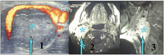
Figure 1. Evidence of tumor mass between the internal and external carotids, presented by the echography with duplex scan of the carotid arteries. Evidence of tumor mass through magnetic resonance imaging in profile and axial cut. - * 1,2 and 3 represent the tumor mass
Through nuclear magnetic resonance imaging a solid and expanding lesion became evident, located in the right parapharyngeal space, measuring 4.6 centimeters in its highest diameter, presenting highly-defined outline forms, lightly lobate, discreet hyper-signal in T2 and hypo-signal in T1 and high gadolinium enhance (Figure 1).
After the clinical suspicion of carotid body tumor, which has been ratified by complementary exams, surgery has been indicated. The surgical team was made of one otorhinolaryngologist, one head and neck surgeon and one vascular surgeon.
Surgery description:The patient was placed at dorsal decubitus position with head hyper-extension in order to provide higher maneuver possibility, if necessary. The Conley incision has been chosen, in "y" shape, in order to obtain high surgical access, thus providing high exposition of sternocleidomastoid muscle and producing three flaps: a cranial, a lateral and a medial one, and each one of them were fixed to the surgery field.
The initial objective of the mass handling was to make the surgery field bloodless, through the blood vessels ligation. After that procedure, the jugular-carotid fascia was accessed, thus initially liberating the external carotid artery and next, the internal carotid artery, thus providing conditions for the total liberation of tumor mass. Once the vagus nerve was incorporated to this mass, it needed to be resected in conjunction. All skull nerve pairs have been preserved, such as hypoglossal, spinal, gloss pharyngeal and the cervical and marginal branches of the facial nerve. The internal jugular veins have also been preserved (Figure 2).
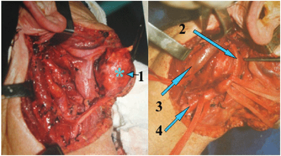
Figure 2. Presentation of tumor mass and cervical vessel (internal jugular vein and internal carotid artery)<}0{> {0><}0{>-1- tumor mass, 2- internal carotid artery, 3 - internal jugular vein and 4 - common carotid artery.
The material removed from the surgery was forwarded to anatomo-pathologic clinic for freezing analysis and paraffin embedding. A "suctor" was placed for surgery cavity aspiration and the stitches were performed into two plans, the subcutaneous and the skin. During the post-operative phase, antibiotics therapy, analgesic medication and the "suctor" were kept.
The histological result was compatible with benign paraganglion, so the immune-histochemical exam was recommended. The result of such exam, for characterization of tumor origin through the S-100 protein, cromogranine A and sinaptophyzine, was positive in the neoplasia cells, which confirmed paraganglion (Figure 3). The patient was hospitalized for 6 days and was then discharged in good clinical conditions.
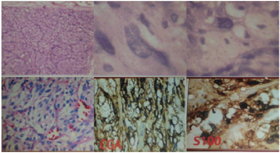
Figure 3. Presentation of stained histological cuts, superiorly, by hematoxiline and eosine and, inferiorly, by cromogranine, and by protein S100.
The tumor belonged to group II, once it was firmly adhered to the arteries, making its resection difficult(13). Even though the surgery team was prepared for the placing of a by pass in any of the arteries, that was not necessary. Such procedure has already become common when its performance is necessary and is performed by many scholars and professionals who deal with this kind of tumor(2), thus obtaining a high reduction of morbidity and mortality.
The carotid body tumors are benign according to a consensus of many authors, but even so they need to be surgically treated(10), once they may present local symptoms such as pain, compression and distance reflex symptoms, such as pain, pruritus, plenitude, dysphalgia and dysphonia. The fact that it may become malign is mentioned in the literature(7). In order to avoid such complications and once the patient already presented signs and symptoms of tumor affection, the elective procedure really was the surgery(8,14).
The anatom-pathological analysis through freezing just revealed that the lesion was not malignant. The paraffin study showed it was a lesion which was compatible with paraganglion, and because it was not conclusive the immune-histochemical analysis was required, which ratified the anatomo-pathological finding.
The symptomatology presented by the patient agrees on other authors' descriptions, thus being the most frequently found ones(4,7,8).
The patient followed the social-demographic profile described by this kind of tumor: she was female, 60 years old. There is a consensus in the literature that the tumors with sporadic appearance occur at 55 years of age and the hereditary ones occur in young patients and at 26(5,6). The tumor in our study was unilateral, which is the most frequently found one.
For diagnosis, the magnetic resonance imaging and echography with duplex scan of the carotid arteries were used; the selective arteriography was not performed, once the diagnostic hypothesis had already been established and it was not available at that moment. He have not performed the pre-operation embolism, once the literature is very clear demonstrating it is little relevant.
At such tumors' resection, eventually there is involvement of skull pairs, which may present some kind of affection or the total loss of its function. In our case, there was an involvement of a vagus nerve segment with the tumor, so we had to resect it in a segmental way, thus resulting on a partial affection of the pharyngeal and laryngeal motor functions. The patient got used to this new situation, and needed phonotherapy.
When no treatment is used with the carotid body tumor, its mortality may reach 30%(10). Nowadays, the elective treatment is the surgery(4,9,10,11). Radiotherapy does not offer therapeutic aid for such tumors, unless there are residual tumor cells(4,9) originated from little safety margin of surgical resection. Some authors use embolism, but they do not use it. Other authors report the eventual need of artery resection and its interposition with other homologous or autologous materials(4). Surgical patients must be followed during a long period of time, once they may have metastatic diseases which may become evident in 10 or 20 years' time(3,12).
In our case, we did not consider embolism necessary, once the tumor was very delimited, respecting the arterial structures, as well as the interposition with other materials was not necessary.
The previous symptomatology which was presented by the patient disappeared after the surgery, with the exception of a dysphonic clinical picture, which has been caused by segmental section of the vagus nerve demonstrated through the laryngoscopy, the positioning of the right vocal cord in a paramedian situation.
CONCLUSIONThe carotid paraganglion is a rare tumor and its treatment is surgical. Due to its high anatomo-surgical complexity, it requires: criterial clinical evaluation, the need to know the differential diagnosis and to establish the definitive one, and the participation of several professionals from different medical specialties.
REFERENCES1. Gardner E, Gray DJ, O'rahilly, R. Anatomia 4a Ed. Rio de Janeiro: Guanabara Kogan; 1988 p. 685.
2. Galvão ARJr, Sartini AL, Machado MC, Mattioli FM, Ribas MH, Fava AS. Bilateral carotid body paraganglioma. Rev Bras Otorrinolaringol 2004; 70(4):573-6.
3. Muhm M, Polterauer P, Gstottner W, Temmela A, Richling B, Undt G, Nierdele B, Staudacher M, Ehringer H. Diagnostic and therapeutic approaches to carotid body tumors. Review of 24 patients. Arch. Surg 1997; 132:279-84.
4. França LHG, Bredt CG, Vedolin A, Back LA, Stahlke Jr. HJ. Tratamento cirúrgico do tumor do corpo carotídeo: J Vasc Br 2003; 2(3):171-5.
5. Gardner P, Dalsing M, Weisberg E, Sawchuk A, Miyamoto R. Carotid body tumors, inheritance and a high incidence of associated cervical paragangliomas. J Surg 1996; 172:196-9.
6. Rabl H, Friehs I, Gutschi S, Pascher O, Koch G. Diagnosis and treatment of carotid body tumors. Thorac Cardiovasc Surg 1993; 41:340-3.
7. Schmind C, Tjan T, Mollhoff T, Schober O, Scheld HH. Recurrent bilateral carotid body tumors. A case report on a "typical" course of a rare disease. Thorac Cardiovasc Surg 1995; 43:296-8.
8. Vedolim AC, Schmitt CMA, Bredt CFG, Barros MB, França LHG, Stahlke Jr HJ.Tumor do corpo carotídeo: análise de 14 casos. RBM Rev Bras Med 2003; 60(5):267-70.
9. Souza AA, Pereyra WJF, Santos LS, Marques JAP, Carvalho GTC. Tumores do corpo carotídeo: Arq Neuropsiquiatr 2000; 58:324-9.
10. Kaman L, Singh R, Aggarwal R, Kumar R, Behera A, Katariya RN. Diagnostic and therapeutic approaches to carotid body tumors: report of three cases and review of the literature. Aust NZJ Surg 1999; 69(12):852-5.
11. Matticari S, Credi, G, Pratesi C, Bertini D. Diagnosis and surgical treatment of the carotid body tumors. J Cardiovasc Surg 1995; 36:233-9.
12. Seabrook GR, Towne JB. Doença vascular cerebral nãoaterosclerótica. In: Haimovici, H. Cirurgia Vascular: Princípios e Técnicas, 4a ed., Rio de Janeiro, Di-Livros; p 975-8, 1999.
13. Shamblin WR, Remine WH, Sheps SG, Harrison Jr EG. Carotid body tumor: chemodectoma. Am J Surg 1971; 122:732-43.
14. Silva ES, Tozzi FL, Paiva FHM, Sukys GA. Bilateral carotid body paraganglioma: Rev. Paul Med 2000; 118(1):13-6.
15. Lack EE, Perez-Atayde AR, Young JB. Carotid body hyperplasia in cystic fibrosis and cyanotic heart disease. A combined morphometric, ultrastructure, and biochemical study. American Journal of Pathology 1985; 119:301-4.
1. Ph.D. in DSB by USP/SP (SaffF the Hospital Odontomed Visiting Professor of scientific methodology of the School of Nursing, Itajubá/MG)
2. Head and Neck Surgeon, and Otorhinolaryngologist by INCA/RJ, PUC/RJ and Centre Oscar Lambret-France. (Staff of the Hospital of Oncology in Varginha, MG and Clinic of San Lucas-Caxambu/MG.)
3. Scholar, School of Medicine Itajubá (Scholar, School of Medicine Itajubá)
4. Ph.D. in DSB by USP/SP. (Full Professor of the Pontifical Catholic University of São Paulo.)
Institution: Odontomed Hospital.
Dr Francisco Sales de Almeida
Address to corresponência: Rua Major Belo Lisbon, 88 - Centre - Itajubá/MG - CEP: 37500-016 - Phone: (035) 3621-2000 - E-mail: fsalesdr@sulminas.com.br
Work submitted on 23/7/2006 12:35:06 and applied the definition on 14/11/2006 03:23:09.

