INTRODUCTION Foreign body ingestion is a common problem (1). Mostly it is accidental but sometimes individuals ingest certain materials to conceal it. The most frequently swallowed foreign bodies in children include coins, metallic foreign bodies (parts of playing objects) and while meat bone (chicken bone/fish bone/mutton/buff) are common in adults and elderly (2,3,4,5,6). Children between the ages of one and three years were the most commonly affected (7). Foreign bodies frequently occur in the cricopharyngeal and oesophageal regions (8). Most of the foreign body which have gone beyond esophagus will pass uneventfully through the intestinal tract (1). Foreign bodies in upper digestive tract whether blunt or sharp should be considered as an emergency to reduce the associated complications. If foreign bodies are not removed on time, it can cause intramural perforation, subacute mediastinitis, aortoesophageal fistula (9), tracheoesophageal fistula, and long-term residual injury to the esophagus (10). This study was done to identify the type and site of foreign body ingestion in both children and adults.
MATERIALS AND METHODSA retrospective analysis of 163 cases of suspected foreign body ingestion were done in patients admitted in ENT& Head and Neck Surgery department of TU Teaching Hospital in between April 2004 and October 2006 (2 ½ years). All age groups with suspicion of foreign bodies were included. Age less than 12 years was included in children while age more or equal to 13 years was included in adults. Informed consent was taken from all patients in order to participate in this study and the work was approved by local ethical committee. In all cases, x-ray soft tissue neck lateral and x-ray chest anteroposterior view done along with other preoperative investigations were done. In all patients rigid oesophagoscopy or pharyngoscopy under general anaesthesia were done to remove foreign bodies. While in two cases flexible endoscopies were done.
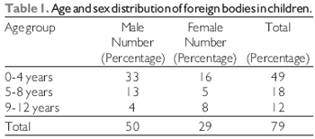
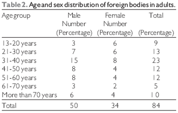
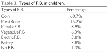
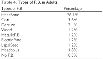
RESULTS There were 163 cases of foreign body ingestion, out of which 48.5% were children and 51.5% adults. Foreign bodies were common in 0-4 year age group in children where as in adults it was common in 31-40 years age group (Table 1, 2). In children, the most common foreign bodies were coin (60.7%), meat bone 15.2%, and metallic foreign bodies 8.9% (Table-3). The most common site of all these F.B. was at cricopharyngeal junction (51.9%), followed by oesophagus (46.8%). In contrast in adults the most common foreign bodies were meat bone (76.1%) followed by coin (3.6%) and denture (2.4%) (Table-4).The foreign bodies were more common (29.8%) in 30-40 years age group. The most common site in adults was oesophagus (62.0%) followed by cricopharyngeal junction (25.0%) and pyriform sinus (4.7%). There were 8.3% of adults and 1.2% of children in whom foreign bodies were not present. There was no mortality noted during entire period of this study.
DISCUSSIONForeign body ingestion is a common occurrence and carries significant morbidity and mortality. The peak age in children is between 6 months to 3 years (7). Our study showed that foreign bodies were common in 0-4 year age group in children where as in adults it was common in 31- 40 years age group. On analyzing 163 cases of ingested foreign body we found almost equal percentage in children and adults/elderly. However, Foreign body were common (29.8%) in 30-40 years age group.
Most common foreign bodies in pediatric age group are coins (2,3,4,5,6), but meat bone, marbles, safety pins, button, batteries and screws are also reported. Our study also revealed coins (60.7%) to be the most common foreign body in children followed by meat bone (15.2%) and metallic F.B. (8.9%).
In old age ingestion of a bolus is common occurrence specially elders who are edentulous who cannot chew properly, particularly food like meat and swallow it as a whole. Moreover elderly patient most of the times have other underlying pathology which needs to be screened.
F.B. can be impacted in the pharynx and oesophagus because of their shape, size and anatomical narrow segments. More adults than children tend to have impaction of bones in the pharynx and oesophagus. The oesophagus is a passive and unadaptable organ and its peristalsis is not strong enough to prevent its retaining certain types of swallowed objects (9). Meat bones are common in an adult which is similar to our study. Our study also showed coins and denture as a common foreign body in adults. The most frequent location was the oesophagus and cricopharyngeal junction. However there are several studies done showing cricopharyngeal and oesophageal region to be the common site (2,8).
Removal of foreign body is not an easy task. It needs proper instruments and skill (2). Any F.B. that is large and impacted or any sharp F. B. should be removed immediately (1). Blunt F.B. can be usually removed safely by rigid endoscope (1).In the recent past, flexible endoscope has been advised for oesophageal F.B., where success rate is 76-95% and 0% morbidity and mortality (10). There is a significant risk of causing laceration and perforation of oesopghagus while removing sharp F.B. with flexible endoscope. These can be avoided by using rigid oesophagoscope.
In all of our patients rigid oesophagoscopy or pharyngoscopy under general anesthesia were done to remove foreign bodies. While in two cases, flexible endoscopies were done and in four cases foreign body were advanced into the stomach. There were 8.3% of adults and 1.2% of children in whom foreign bodies were not present two cases of children had associated oesophagial stricture.
Sharp F.B. is frequently associated with serious complications. If they are not removed at the earliest, they can cause erosion, perforation, abscess or mediastinitis (3). The incidence of such complications occurs even after the removal of F.B which is often due to anesthesia, or due to delayed presentation. There were no morbidity and mortality noted in our case series. However other studies showed complications like oesophageal perforation, oesophagoaortic fistula, empyema thoracis, mediastinitis and lung abscess (3,8).
CONCLUSIONBlunt foreign bodies were common in children while sharp foreign bodies were in adults. Early removal of these foreign bodies must be considered to reduce the risk of complications. It is better to prevent children by not allowing them to play with coins/metallic foreign bodies/safety pins etc. Parents/caretaker should be educated to take their children to the hospital even there is a suspicion of foreign body ingestion. Even if there is a not a clear cut history of foreign body ingestion and if you suspect, you should not neglect it.
REFERENCES1. Shivakumar AM, Naik AS, Prashanth KB, Yogesh BS, Hongal GF. Foreign body in upper digestive tract. Indian J Pediatr 2004;71:689-693.
2. Nandi P, Ong GB. Foreign body in the oesophagus: review of 2394 cases. Br J Surg 1978; 65:5-9.
3. Guitron A, Adalid R, Huerta F, Macias M, Sanchez Navarrete M, Nares J.Extraction of foreign bodies in the esophagus. Experience in 215 cases. Rev Gastroenterol Mex 1996; 61:19-26.
4. Yang CY. The management of ingested foreign bodies in the upper digestive tract: aretrospective study of 49 cases. Singapore Med J 1991; 32:312-5.
5. Morales-Angulo C, Rodriguez Iglesias J, MazonGutierrez A, Gomez Castellano R, Rama J. Acta Otorrinolaringol Esp 1998; 49:644-6.
6. Nayak SR,Kirtane MV,Shah AK,KArnik PP. Foreign bodies in the cricopharyngeal region and oesophagus (a review of 226 cases). J Postgrad Med 1984; 30:214-8.
7. Brown DA, Clark CM. Inhaled foreign bodies in children. Med J Aust 1983; 2:322-6.
8. Vella, E. E. and Booth, P. J.: Foreign bodies in the oesophagus. Brit. Med. J 1965; 2:1042-1043.
9. Wilson RT, Dean PJ, Lewis M. Aortoesophageal fistula due to a foreign body. Gastrointest Endosc 1987; 33: 448-50.
10. Jona JZ, Glicklich M, Cohen RD. The contraindications for blind esophageal bouginage for coin ingestion in children. J Pediatr Surg 1988; 23:328-30. Adhikari P Arq.
1 Dr. M.S. First Year Resident, Department of ENT & Head and Neck Surgery, T.U. Teaching Hospital, Kathmandu, Nepal.
2 Dr. M.S. Second Year Resident, Department of ENT & Head and Neck Surgery, T.U. Teaching Hospital, Kathmandu, Nepal.
3 Associate Professor. Associate Professor, Department of ENT & Head and Neck Surgery, T.U. Teaching Hospital, Kathmandu, Nepal.
4 Professor, Department of ENT & Head and Neck Surgery, T.U. Teaching Hospital, Kathmandu, Nepal.
Departmetn of ENT & Head and Neck Surgery, T.U. Teaching Hospital, Kathmandu, Nepal.
Dr. Prakash Adhikari, M.B.B.S., M.S. First Year Resident, Departmetn of ENT & Head and Neck Surgery, T.U. Teaching Hospital, Kathmandu, Nepal.
Tel:+977-1-4414191,Fax:+977-1-4414191. E-mail: prakash_ooz@hotmail.com.
No financial support was avaliable.
Este artigo foi submetido no SGP (Sistema de Gestão de Publicações) da R@IO em 4 de maio de 2007 . Cod. 247. Artigo aceito em 18 de junho de 2007.