INTRODUCTIONFine needle aspiration cytology (FNAC) has gained popularity as a rapid cost-effective, relatively painless, precise and effective means to diagnose thyroid and salivary gland lesions and other masses in head & neck. However its use in the diagnosis of oral & oropharyngeal lesions is not being commonly practiced. Almost all of the oral & oropharyngeal lesions are subjected to punch biopsy in many centers.
Physicians in developing countries have been found to be dependent upon histopathology reports of the neoplasms even though FNAC is comparatively very much cost effective. Whereas, biopsy is time taking, expensive, more painful, producing unwanted bleeding at times and needs more time for its processing and reporting leading to delay in the planning of definitive treatment. More over many a time a surgeon does not like an encapsulated tumor to be breached.
Very few studies have been done to explore the potential of FNAC for the diagnoses of oral and oropharyngeal lesions. This study was an attempt to find out the efficacy and reliability of FNAC in the diagnosis of Oral & Oropharyngeal lesions.
PATIENTS AND METHODSThis was a hospital based prospective blind study conducted in the Department of Otolaryngology Head & Neck Surgery and the Department of Pathology, T. U. Teaching Hospital, Katmandu, Nepal. All clinically diagnosed cases of Oral & Oropharyngeal tumors of all ages and both sexes attending the outpatient department during the above mentioned period were taken up for the study. A total of 52 aspirations were carried out. Among which three patients were excluded from the study, two because there were no histopathology report for correlation and one because there was insufficient material for cytological diagnosis.
All aspirations were carried out in the ENT outpatient department. The procedure was well explained to the patients and due consents were taken from all of them. All patients received local anesthesia in the form of Lignocaine spray or lignocaine viscous gargle to avoid gag reflex and pain. The samples were taken with the patients in a supine or sitting position with a head support. Visibility was enhanced by using a head mirror. The aspirations were performed with a 23 gauged needle attached to a 10 ml disposable syringe. The needle was introduced into the target and suction applied by retracting the syringe plunger to the 1-2 ml mark. The needle was moved back & forth four to five times in the same plane to ensure minimal bleeding. Four samples were collected. Two slides were air dried for Giemsa staining and two were fixed in 95% alcohol for Papanicolaou stain.
Inadequate aspirates that were not available for repeat aspirations were excluded from the study. FNAC report without histological correlation was also excluded from the study. The patients were then subjected to biopsy immediately after aspiration with a punch biopsy forceps and the specimen fixed in 10% formalin. Some of the patients underwent surgery at a later date and the surgical specimen was subjected to histopathological examinations. The FNAC was reported by a single consultant cytopathologist who was not aware of the histopathological examination report. The HPE reporting was done by a consultant pathologist who was not aware of the FNAC report.
FNAC and HPE reports of these patients were reviewed to find out the accuracy of FNAC with the HPE report. Data was analyzed according to the method of Galen and Gambino. Absolute sensitivity was the positivity of FNA when malignancy was present, that is, the fraction of patients with histologically documented malignant neoplasms for which the test was unequivocally positive. Complete sensitivity was the fraction of the patients in whom histologically malignant neoplasm was detected by FNA either as unequivocally positive or suspect.The false negative rate was the fraction of patients with histologically malignant disease in whom FNA failed to detect any abnormality. Specificity measured the ability of the test to have unequivocally benign results when malignancy was absent.
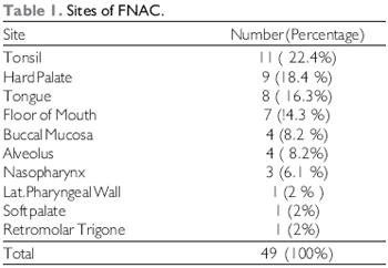
RESULTSForty nine patients who had both FNAC and HPE reports were taken for analysis of data. Among the total patients included in the study there were 24 male and 25 female patients. Age of the patients ranged from 14 months to 84 years with a mean age of 46.9. The maximum number of patients were in the 40 - 59 years of age (22 patients) followed by 20 - 39 yrs. (14 patients). A total of ten sites were aspirated from the oral and pharyngeal regions. Maximum (11) aspirates were from tonsils followed by hard palate (9), tongue (8), floor of mouth (7) etc (Table 1).
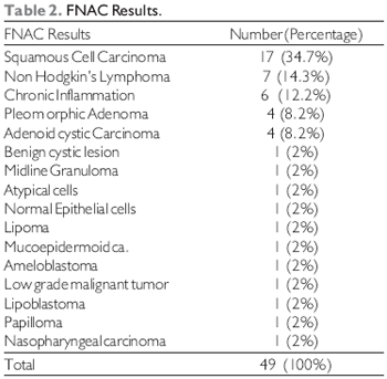
FNAC ResultsThe FNAC results revealed 32 malignant and 16 benign results with one aspirate showing atypical cells where biopsy was advised to rule out malignancy. Among the malignant lesions squamous cell carcinoma was seen in seventeen patients followed by Non Hodgkins Lymphoma in seven smears. Among the benign lesions chronic inflammation was seen in six patients followed by pleomorphic adenoma in 4 patients (Table 2).
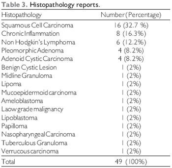
Histopathology Reports Histopathology of the subsequent punch biopsy or surgically excised specimen showed malignant lesions in 31(63.3%) and benign in 18(36.7%) patients. (Table 3).
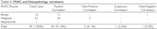
Correlation of FNAC with Histopathology ReportsIt was found that fifteen benign lesions and thirty malignant tumors reported in FNAC correlated with the histopathology results. One sample where FNAC had reported as chronic inflammatory cells turned out to be tuberculous granuloma histopathologically. But there was one false positive result in the benign group where aspiration from the alveolus showed pleomorphic adenoma but the histopathology showed chronic inflammatory cells only.
Likewise there were two false positives where FNAC showed squamous cell carcinoma and NHL, but histopathology showed chronic inflammatory cells and normal lymphoid tissue respectively. And in one case which was reported as atypical cells was later reported as Verrucous carcinoma on histopathology (Table 4).
Site specific correlationThere was one false positive each in nasopharynx and tonsil which was later seen to be chronic inflammation and normal epithelial cells and a false negative in tongue which was tuberculous granuloma and a suspicious lesion in the floor of mouth which turned out to be verrucous carcinoma.
Negative / Positive correlationThere were two false positive results and one false negative result. The false positives were of the aspirations carried out from the tonsil and nasopharynx and the false negative aspiration was carried out from the tongue. One patient with cytodiagnosis of NHL from the tonsil was reported to have chronic inflammation and the other cytodiagnosis of Squamous cell carcinoma from the nasopharynx on histological examination was seen to have chronic inflammation.
In one patient with an ulcerative lesion of tongue was found to have normal epithelial cells in FNAC showed to have tuberculous granuloma in histopathology. In another case even though atypical cells was found in FNAC, it could not confirm the diagnosis of Verrucous carcinoma which was reported histopathologically.
Data analysisAs a whole, it was found that the absolute sensitivity was 97.83%, complete sensitivity 97.87% and Specificity 88.35% with Positive predictive value 93.93% and negative predictive value 93.75%. Considering the malignant tumors alone, the Absolute as well as Complete Sensitivity was found to be 100% with Specificity to be 88.24%. As far as the benign tumors were concerned, the Absolute and the Complete Sensitivity was found to be 93.75% with Specificity to be 88.24%.
DISCUSSIONBiopsy has been the established diagnostic procedure to confirm the diagnosis of oral and oropharyngeal lesions. But sometimes surgeons do not like to breach the overlying epithelium of the lesions before coming to a probable diagnosis. Moreover, in developing countries, it takes nearly two weeks to get the histopathology report whereas FNAC report can be obtained within a couple of days. Hence keeping these points in mind, this study was carried out to explore the potential of FNAC as an alternative diagnostic procedure.
There are some difficulties in performing FNAC from Oral & Oropharyngeal tumors in the dept. of Pathology due to the technical reasons like lack of proper light, positioning of the patients and difficult anatomic locations etc.
In the current study a total of 52 aspirations were performed out of which , three had to be excluded , one because of inadequate sampling and two because the patients were not available for biopsy. The age of the patients ranged from 14months to 84 years. The youngest one was a case of Lipoblastoma of cheek and the oldest one a case of NHL.
The accuracy of FNAC varies widely both in published literature as well as in the community (1). Training and ability in specimen interpretation is important (2). Studies have shown that the majority of missed diagnosis are due to problems in sampling and specimen preparation (3, 4). In this study the sampling has been adequate in all cases except in one. Of course repeat aspiration had to be performed in two cases for adequacy.
In this study there was only one false negative report which turned out to be a tuberculous granuloma of tongue where the cytopathological diagnosis was normal epithelial cells. Likewise there were two false positives on histological correlation. One was a FNAC from nasopharynx which on histopathological examination showed only chronic inflammatory cells. The other aspirate was from tonsil which was reported as NHL but showed only chronic inflammatory cells with normal lymphoid tissue on histopathological examination. Interestingly these patients also had lymph nodes that on aspiration were diagnosed as metastatic squamous cell carcinoma and NHL respectively. Hence FNAC can be a better option sometimes in cases of deeply sited lesions like tonsils where biopsy might be superficial and miss the targeted tissue.
There was another case where suspicion of atypia was raised on FNAC which later proved to be verrucous carcinoma. Very few studies have been done to find out the accuracy of FNAC in oral and Oropharyngeal tumors. Regarding the role of FNAC, SCHER et al has emphasized the positive predictive value of 100% (6).
The diagnostic accuracy of FNAC in the current study for the same sites was 93.75%,whereas absolute sensitivity was 97.83%,complete sensitivity 97.87% and specificity 88.35% with an overall accuracy of 93.75% which is comparable to the study done by SHAH et al who had an absolute sensitivity of 93.3%, complete sensitivity of 93.9% and specificity of 85.7% (7).
Insufficient sampling and diagnostic dilemmas can be eliminated when smears are stained and evaluated immediately after aspirations with repetition of the procedure until adequate sample is obtained and the reported smear inadequacy of 2 - 10% can be minimized. In this study inadequate sampling was seen in only one patient.
SEETHARAMAN et al. in their study found that in Oral squamous cell carcinoma FNAC result was true positive in 92.85% and 7-14% was false negative where as in cases of leukoplakia , overall positive correlation was found in only 33.33%.So he concluded that FNAC can be used as a reliable diagnostic test for oral squamous cell carcinoma but its use in oral leukoplakia is of limited value (5).
GUNHAN et al. studied the efficacy of FNAC of oral cavity and jaw bone lesions in 102 cases. He found that 13 out of 15 histologically malignant lesions and 71 out of 87 histologically benign lesions were cytologically diagnosed correctly. Hence he concluded that the overall accuracy of FNAC in oral cavity and jaw bones is high (8).
SCHER et al. in his study of the role of FNAC in the diagnosis of lesions from Oral cavity, oropharynx, and nasopharynx found no false positive diagnosis in cases of malignant lesions. In 16% of the cases FNA was unsatisfactory and hence he emphasized the need of repeating the FNA or recommending biopsies in negative and unsatisfactory FNA when clinically indicated to assure the accuracy in diagnosis. There were no complications resulting from the FNA (6).
In recent years the existing controversy on the diagnostic accuracy of FNAC in hematologic malignancies has been cleared and a significant number of studies have shown that not only is the cytologic assessment of these tumors possible, it is also highly accurate. (9,10).Similarly in this study there were 7 aspirates diagnosed as Non Hodgkin's Lymphoma (NHL) but on histopathological examination one of them turned out to be only chronic inflammation but the excised lymph node of the same patient was reported as NHL and hence was treated in the line of NHL with satisfactory results. Hence it would not be unfair for this study to claim a diagnostic accuracy of 100% for hematologic malignancies of the oral cavity and oropharynx.
Of the total 11aspirates from tonsils 3 had benign, 2 had squamous cell carcinoma and 5 had NHL that have correlated well with the histopathology So in this study FNAC has been highly accurate in the diagnosis of tonsil lesions.
Similarly in hard palate lesions all of the 9 aspirates (with 6 benign and 3 malignant) correlated well on histopathology. In case of the total of 8 tongue aspirates, 6 were reported as Squamous cell carcinoma and 2 benign. There was one false negative report where a cytological diagnosis of chronic inflammation was made but on histopathology turned out to be tuberulous granuloma. It can be concluded that FNAC can be a highly accurate diagnostic procedure for malignant tumors. This is probably due to the decreased cohesiveness of cells in malignancies making adequate aspiration easier for cytological interpretation.
Traditionally, authors advocate a biopsy sample of at least 1.5 cm long to avoid confusion in histopathological reporting.(11) Obtaining biopsy specimen of this size from oral & oropharyngeal sites cannot be always possible and can be very painful to the patient and there is always a greater risk of bleeding.
This study illustrates the variety of lesions benign and malignant found in oral cavity and oropharynx and the power of FNAC in diagnosing the spectrum of different lesions. Moreover conventional biopsy from tonsil can be fraught with severe bleeding .It was found that FNAC of the tonsil provided a safe and effective means of diagnosing deeply sited lesions like Lymphoma or carcinoma which could be missed if a superficial biopsy is taken.
CONCLUSIONFNAC has been highly accurate for the diagnosis of lesions of the tonsils , hard palate and floor of mouth. It was also seen that the study proved to be highly accurate for the malignant lesions which can be of great help in early planning of the definitive course of management.
BIBLIOGRAPHY1. Giard RW, Hermans J. The value of aspiration cytologic examination of the breast: a statistical review of the medical literature. Cancer, 1992:69:2104-2110.
2. Zarbo RJ, Howanitz PJ, Bachner P. Interinstitutional comparison of performance in breast fine needle aspiration cytology: a Q - probe quality indicator study. Arch Pathol Lab Med, 1991:115:743-750
3. Lee KR, Foster RS, Papillo JI. Fine needle aspiration of breast: importance of the aspirator. Acta Cytol, 1987;31:281-284.
4. Cohen MB, Rodgers C, Hales MS et al. The importance of training and experience in the interpretation of fine needle aspirates of the breast: analysis by ROC curves. Arch Pathol LabMed 1987;111:518-520.
5. AU Seetharam SS. Ramchandran CR. Fine needle Aspiration cytology as a diagnostic test for oral squamous cell carcinoma. Oral Diseases. 4(3):180-6, 1998 Sep.
6. Scher RL. Oostingh PE. Levine PA. Cantrell RW. Feldman PS. Role of fine needle aspiration in the diagnosis of lesions of the oral cavity, oropharynx and nasopharynx. Cancer 62(12):2602-6, 1988 Dec 15.
7. Shah SB, Singer SI, Liberman E Ljung BM, Transmucosal fine needle aspiration/Diagnosis of Intraoral and Intrapharyngeal lesions: Laryngoscope, 109, Aug. 1999. 1232-1237.
8. Gunhan O. Dogan N. Celasun B. Sengun O. Onder T.Finci R. Fine needle aspiration cytology of oral cavity and jaw bone lesions. A report of 102 cases. Acta Cytologica. 37(2);135-41, 1993 Mar-Apr.
9. Sneige N. Diagnosis of lymphoma and reactive lymphoid hyperplasia by immunohistochemical analysis of fine needle aspiration biopsy. Diag Cytopathol, 1990;6:39-43.
10. Daskalopoulou D, Harhalakis N , Maouni N , Markidou S. Fine needle aspiration cytology of non hodgkin's lymphoma. A morphologic and immunophenotypic study. Acta Cytol, 1995;39:180-6.
11. Scully C. Clinical diagnostic methods for the detection of premalignant and early malignant oral lesions. Community Dental Health, 1993;10:43-52.
1. Dr. Resident, Department of ENT and Head and Neck Surgery, T. U. Teaching Hospital, Kathmandu, Nepal.
2. Professor, Department of ENT and Head and Neck Surgery, T. U. Teaching Hospital, Kathmandu, Nepal.
3. Professor and HOD, Department of Pathology, T. U. Teaching Hospital, Kathmandu, Nepal.
4. Dr. Associate Professor, Department of ENT and Head and Neck Surgery, T. U. Teaching Hospital, Kathmandu, Nepal.
5. Dr. Associate Professor, Department of ENT and Head and Neck Surgery, T. U. Teaching Hospital, Kathmandu, Nepal.
6. Dr. M.S.Resident, Department of ENT and Head and Neck Surgery, T. U. Teaching Hospital, Kathmandu, Nepal.
Department of ENT, Head & Neck Surgery, T. U. Teaching Hospital, Kathmandu, Nepal. Prof. Bimal Kumar Sinha, Department of ENT, Head & Neck Surgery, T.U.Teaching Hospital, Kathmandu, Nepal
This article was submitted in SGP (System of Administration of Publications) of R@IO on February 23, 2008. Cod. 421. Article accepts on March 14, 2008.