INTRODUCTIONBeing essential to the practice of modern histopathology, the immunohistochemical technology has been considerably developing since its introduction in early 1940's. This method is based on the demonstration of the antigen-antibody reaction with the possibility of its identification at optical microscopy, which may be done through immunofluorescence or through immunoenzymatic methods. In recent years, the role of immunohistochemistry as a diagnostic method has been changing from assistant technique to main technique, which may determine the best therapy approach and prognosis.
Several studies involving immnunohistochemical methods to evaluate the epidermis of the external acoustic meatus were performed with very different objectives. Through immunohistochemistry, initially used to diagnose tumors, the epithelial migration mechanisms from skin to meatus, the local immune response, the expression pattern of intermediate filaments, the presence of cytokines and other antigens, assessment of new proposals to treat diseases which affect the meatus, as well as the cholesteatoma, among others, may also be studied.
With a deeper knowledge of the immunohistochemical patterns of the epidermis of the external auditory canal, there may be a higher understanding of its relations with the genesis of the several diseases which affect it, as for example, the cholesteatoma, besides contributing to choose the best treatment and to establish the prognosis for each disease.
One of the most accepted theories for the genesis of cholesteatoma is the migration of epidermis of external auditory canal to the interior of the medium ear cavity through a perforation of the tympanic membrane. However, there are still several questions among authors, for example why such migration occurs, which usually has centripetal route in the tympanic membrane and lateral one in the external auditory canal, why it occurs only in some people, what makes the cholesteatoma more or less aggressive, among other questions. For such reason, it is necessary to search for a better understanding of the immunohistochemical characteristics of the epidermis of the external auditory canal in order to try to better understand what differently happens in this tissue.
The method chosen to reach the proposed objectives was the systematic literature review, which is an important medical resource, based on evidences which consist of a way to summarize the results of the researches related to a problem or to a specific issue.
To analyze the studies involving the immunohistochemical standards of the epidermis of the external auditory canal, a review of all identifiable articles which were published until 2007 in national or international journals was done about the immunohistochemical methods used in skin sample of the external auditory canal (EAC), the immunohistochemical patterns of the epidermis of the normal meatus and the application of such methods in the several diseases which affect it.
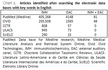
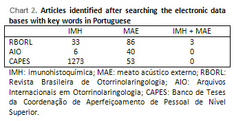
In order to do this survey, the electronic data base (MEDLINE), OVID, Revisões Sistemáticas da Colaboração Cochrane (CCTR), Literatura Latino-Americana e do Caribe em Ciências da Saúde (LILACS) and Scientific Eletronic Library Online (SciELO) were used, in addition to Brazilian publications Revista Brasileira de Otorrinolaringologia (RBORL) and Arquivos Internacionais de Otorrinolaringologia (AIO) and of the thesis bank of Coordenação de Aperfeiçoamento de Pessoal de Nível Superior (CAPES).
For the accurate selection of the key words, the remissive index of three journals indexed in MEDLINE with impact factor higher than 1.0 were analyzed, which had articles related to the subject of this study. They were: the Laryngoscope (Qualis A; impact factor in 2005: 1.617), with the article by Vennix et al (1996), International Journal of Molecular Medicine (Qualis A; impact factor in 2005: 2.090), with the article by Naim et al (2004) and Microscopy Research and Technique (Qualis A; impact factor in 2005: 2.323), with the article by Sirigu et al (1997). After the analysis of such articles, the key words immunohistochemistry and external auditory canal were selected, once they are the ones which are closely related with the objectives of the investigation and the ones which identified studies the most. For the investigation in the Revista Brasileira de Otorrinolaringologia and in the Arquivos Internacionais de Otorrinolaringologia the words were used in Portuguese (imunohistoquímica and meato acústico externo). The terms immunocytochemistry and external acoustic meatus were also used for the investigation, in addition to the combination of all words mentioned, which brought a result of a smaller number of articles.
After such investigation, the bibliographic references of the articles included in this study were also evaluated in order to include studies which were not identified by the investigation in the data base with the key words chosen.
The inclusion criteria of this systematic review were:
-Articles published in indexed journals and identifiable in world literature;
-Thesis or essays identified manually or through data base search;
-Articles whose objectives are directly or indirectly related with the immunohistochemical assessment of the epidermis of the normal or modified external auditory canal;
-Articles whose immunohistochemical method, as well as the antibodies used were described in details.
The exclusion criteria were:
-Articles whose objectives are not directly or indirectly related with the immunohistochemical assessment of the epidermis of the normal or modified external auditory canal;
-Articles whose immunohistochemical method, as well as the antibodies used were not described in details.
-Articles concerning diseases which do not occur in the external auditory canal and with no relation with the epithelium;
-Articles whose antigen used were not directly related to the epithelial tissue or with cholesteatoma;
- Articles published in articles which are difficult to be translated (Japanese, Chinese, German).
The articles included in this study were divided and analyzed per topic:
- Articles which are exclusively about normal external auditory canal;
- Studies which co-relate the epidermis of the external auditory canal with the cholesteatoma;
- Immunohistochemical studies about the epidermis of modified external auditory canal.
This study aimed at reviewing, describing and analyzing the knowledge brought by studies identified in the world literature concerning immunohistochemistry of epidermis of the external auditory canal.
LITERATURE REVIEW
Studies which are exclusively about normal external auditory canalBroekaert, Boedts [1] (1993) performed an immunohistochemical study to investigate the expression pattern of cytokeratins 4, 5, 7, 8, 10, 13, 14, 16, 18 and 19 in fragments of the skin of the external auditory canal, of the epithelial portions and of the mucosa of the tympanic membrane and of the mucosa of the middle ear. The authors observed the expression of CK 5 and 14 in the basal layer and of CK 10 in the suprabasal layers in the epidermis of the tympanic membrane and in the EAC. In the region of the fibrocartilaginous ring, expression of CK 19 was found in the basal layer and of CK 16 was found in the suprabasal layers both in the tympanic membrane and in the osseous part of the EAC. In the other regions of the canal, there was reaction for CK 16 just in the epithelium covering the pilous follicles.
Lepercque et et al [2] (1993), analyzed the distribution of cytokeratins 4, 5, 7, 8, 10, 13, 14, 16, 18, 19 through immunohistochemistry in the epidermis of the external auditory canal and of the tympanic membrane. The external part of the tympanic membrane, made of keratinized pavimentous stratified epithelium, showed a pattern similar to the epidermis, with expression of CK5, CK10 and CK14. In addition to this, a focal expression of CK19 was observed in the inferior region of the fibrocartilaginous ring, in the keratinocyte of the basal layer. A CK16 was predominantly found and located in the suprabasal layers of the epidermis of the region of the tympanic membrane which is near the ring, with gradual reduction of coloring intensity in direction to umbo. In the epithelial samples removed from the cartilaginous portions of the external auditory canal, the expression of CK5, CK10 and CK14 were also observed, which are always present in the epidermis. There was no detection of CK16 and CK19 in this region of the canal. However, in the region closer to the annulus tympanicus, besides the CK which are typical of epithelial tissue, the expression of CK16 and CK19 were also found respectively in the suprabasal and basal layers.
Vennix al [3] (1996) evaluated the expression of cytokeratins in several parts of the external auditory canal in order to use it as characterization parameter of epithelial differentiation. In the cartilaginous part of the EAC, all basal bells were stained with the antibodies for the cytokeratins of basal cells, CK5 and CK14. In the osseous canal such cytokeratins were evidenced not only in the basal layer but also in the suprabasal layers in most samples. Both in the cartilaginous part and in the osseous part of the EAC and in the tympanic membrane positivity was observed in all cells of suprabasal layers for the keratinization marker, CK10. There was no reaction in the skin of the cartilaginous canal with the cell markers associated to proliferation, CK6 and CK 16, neither with the specific antibodies for simple and stratified epithelium, including CK7, CK8, CK17, CK18 and CK19. In the osseous canal CK6 expression was detected in more than two layers of suprabasal cells with involvement of basal cells in some places and of CK16 and CK17 in one or more suprabasal and basal layers, respectively. One CK19 was present just in cell agglomerates in basal layer, in which the CK6 was not detected. In the tympanic membrane, all cells of basal layer were stained by antibodies against CK5 and CK14. In all samples of tympanic membrane, both CK16 and CK17 presented a homogeneous expression pattern in one or more suprabasal cell layers of the ring, of the peripheral portion of the tense part, of the flat part and of umbo area, and the central region of the tense part had variation of expression since a focal pattern to a homogeneous one. The CK19 presented irregular expression pattern of basal cells in the area of the fibrocartilaginous ring, of the flat part, of the umbo region and of the peripheral portion of the tense part. In the central portion of the tense part, the CK19 presented a pattern which was similar do the patterns of CK16 and CK17.
In order to assess the epithelial migration of the epidermis of the external auditory canal, Kakoi et al [4] (1997) analyzed the expression of Ki-67, which is a nuclear and nucleolar antigen closely related to cell proliferation, and the expression of proliferating cell nuclear antigen (PCNA), which is an antigen found in cells which are under division, in samples of skin of tympanic membrane and of the external auditory canal. There was no significant difference in the distribution patterns of the nucleus marked between the PCNA and the Ki-67. Immunoreactivity was demonstrated in the tense part for both antigens in the nucleus of epidermis cells both close to the umbo, mostly in basal cells, and in the region of the fibrocartilaginous ring. Different from the umbo, in the different areas of the ring there was a significant variation both in the number of immunoreactive nucleus and in the distribution patterns. In the inferior quarters of the tympanic membrane, nucleuses with positivity were in higher quantity and more diffuse in the ring, reducing its size close to the skull. In the EAC epidermis there were several marked cells, present in higher quantity in the inferior part of the ring region.
Sanjuan et al [5] (2007), in order to discover if the keratinocyte features of the retroauricular epidermis and of the external auditory canal continue the same before and after its culture, investigated through immunohistochemistry the expression of several cytokeratins in the epithelium of the normal external auditory canal, in the retroauricular skin and in the cells of such regions obtained through culture. The EAC epidermis presented homogeneous expression of CK5 in the basal layer and of CK10 in the suprabasal layers. CK16 expression was not found in any sample. The cells obtained through culture present almost the same expression pattern of the cytokeratins, which shows that the features of the cytoskeleton of keratinocytes of the skin of the canal and retroauricular after culture are kept.
Studies which co-relate the epidermis of the external auditory canal with the cholesteatoma;In all studies in which the immunohistochemical analysis of the epithelium of the external auditory canal was done as control, the studied disease was the cholesteatoma, both in the middle ear and the external ear.
Takahashi, Nakano [6] (1989) investigated the localization of Langerhans cells in the epidermis of the external auditory canal, in the tympanic membrane and in the cholesteatoma of middle ear with the use of immnunohistochemival methods. For this study, the antibody anti-S100 was used, which identifies the S-100 protein, which is present in the cells derived from the neural crest which showed to be an important marker of Langerhans epidermal cells. Few cells were found dispersed in the normal epidermis of the external auditory canal and of the tympanic membrane, just in the stratum spinosum. A relatively higher quantity of reactive Langerhans cells for S-100 protein was found in the cholesteatoma samples, mainly in otorrhea cases. They were present not only in the stratum spinosum but also in the sub-epithelial region.
Van Blitterswijk et al [7] (1989) studied, though immunohistochemical methods, the expression patterns of cytokeratins in five samples of epithelium of the external auditory canal and of the middle ear besides seven samples of cholesteatoma obtained from patients who underwent middle ear surgery. In the epidermis of the external auditory canal positive reactions for CK10 were found in the suprabasal layers. Different from the cholesteatoma samples, no traces of CK 4, 8, 18 and 19 were found.
Broekaert et al [8] (1992) studied the expression of cytokeratins 4, 5, 7, 8, 10, 13, 14, 18 and 19 through immunohistochemistry in the epidermis of the external auditory canal, of the tympanic membrane, mucosa of middle ear and in the epithelium of congenital and acquired cholesteatomas. The authors observed the expression of CK 5 and 14 in the basal layer and of CK 10 in the suprabasal layer. In the region of the fibrocartilaginous ring, as well as in the region of the EAC close to the tympanum, there was CK 16 expression in the suprabasal layers and of CK 19 in the basal layer, which was more observed in the inferior region. The cholesteatoma epithelium, both the acquired one and the congenital one, presented a localization which was similar to the cytokeratins, however it had more positivity.
Schilling et al [9] (1992) investigated through immunohistochemical methods the distribution of two molecular cells of interleukins 1(IL-1), the IL-1-
a and the IL-1-
b, in the normal epidermis of the external auditory canal, in the hyperproliferative epithelium of the cholesteatoma of the middle ear and in the skin of the retro-auricular region. The IL-1 is a cytokine which acts as an autocrine growth factor for normal keratinocytes and is capable of producing bone erosion. Both the epidermis of the external auditory canal and the retroauricular epidermis showed positivity for IL-1-
a and IL-1-
b in similar intensity in all layers of the epidermis. When the IL-1 expressions in the epithelium of the EAC and of the cholesteatoma were compared, the latter was more intense in all layers of the epithelium.
Sasaky, Huang [10] (1994) examined the expression of cytokeratins 13 and 16 in the epithelium of the external auditory canal, of the tympanic membrane and of the cholesteatoma of middle ear through immunofluorescence. In order to perform the immunohistochemical analysis two monoclonal antibodies were used: the antibody K8.12 in order to identify both cytokeratins and the KS-1A3 just for the CK13. In this study, the epidermises of the EAC and of the tympanic membrane were stained by the antibody K8.12 in the suprabasal layers but not by the antibody KS-1A3, suggesting the presence of CK16 in such layer. The same pattern was found in the cholesteatoma of the suprabasal layers in addition to the CK13 expression in the basal layer.
Lee et al [11] (1994) studied the expression patterns of cytokeratins in skin samples of the external auditory canal and of cholesteatoma of middle earobtained by tissue culture. For the immunohistochemical evaluation, the samples were evaluated with the use of monoclonal antibodies for CK4, CK5 + CK8, CK10, CK13, CK14, CK18 and CK19. The specific antibodies for cytokeratins 14 and 19 (usually present both in simples epithelia and stratified epithelia) stained in a consistent way and with the same intensity both tissues, the first stained the cell of the basal and suprabasal layers and the second just the suprabasal layer. The specific antibodies for cytokeratins 4, 10 and 13 (usually present in the suprabasal layers of stratified epithelia) stained both epithelia with more consistent reaction of the two first ones and less consistent one of the last one.
Schilling et al [12] (1996), in order to clarify the proliferation mechanisms of the cholesteatoma epithelium, studied the tenascin and Ki-67 expressions in skin samples of the external auditory canal and of the cholesteatoma of middle ear. Tenascin is an adhesive glycoprotein of the extracellular matrix expressed in epithelium-mesenchymal interactions during embryogenesis. The coloring patterns obtained with the monoclonal antibodies against the tenascin were co-related with the degree of cell proliferation, detected by the monoclonal antibody MIB (Ki-67). In the epidermis of the external auditory canal there was tenascin and MIB-1 expression. The latter presented a stronger reaction with almost continuous coloring pattern with the superior part of the dermis, with deeper positive reaction in the epidermal layers. An intense expression of tenascin was found in the cholesteatoma creating a continuous band in the epithelium-subepithelium junction reaching the deeper regions of the subepithelial conjunctive tissue. The reactivity to MIB-1 was observed not only in the basal and supranasal layers of the cholesteatoma epithelium but also in some histiocytes and stroma fibroblasts.
Bujía et al [13] (1997) examined the expression of the receptor II of Interleukin - 1 (IL-1-R-II) in 10 samples of normal skin of the external auditory canal and 20 samples of cholesteatoma of the middle ear obtained during the surgery. For its identification human monoclonal antibodies anti-IL-1-R-II were used. In the epidermis of the external auditory canal, the IL-1-R-II was found in the keratinocytes of the basal and suprabasal layers, with homogeneous distribution. In the cholesteatoma samples the epithelial localization of the receptor was similar to the meatus skin, but with three times higher positivity. The authors suggest that the presence of IL-1-
a, in the cholesteatoma epithelium associated to the expression of IL-1-R-II indicates that there is an autocrine stimulation system of keratinocytes of cholesteatoma through IL-1.
Chung, Yoon [14] (1998) investigated the presence of cytokeratins in sample cultures of normal epithelium of the external auditory canal and of cholesteatoma of middle ear with the use of immunohistochemical methods, and also investigated the production difference in IL-1-
a, IL-1-
b e IL-8 between both tissues through the ELISA method (Enzime-linked Immunosorbent Assay). The 14 EAC and cholesteatoma skin samples used in such study were obtained during middle ear surgery. For cytokeratin studies monoclonal antibodies anti-CK (AE1/AE3) were used. Both the EAC epithelium and the cholesteatoma one were immunoreactive to (AE1/AE3), but the localization of positive cell layers was not specified in this study. There was a great difference in the concentration of IL-1-
a, IL-1-
b e IL-8 in all cuts. The IL-1-
a had 2.5 times higher positivity in the cholesteatoma in relation to the epidermis of the external auditory canal. The IL-1-
b had twice higher positivity in the cholesteatoma. The IL-8 had three times higher positivity in the cholesteatoma. The authors suggested that the IL-1-
a and the IL-8 present in the epithelium of cholesteatoma are liable to the bone destruction caused by such disease, and that certain substances originated from the subepithelial granulation tissue may stimulate the production of certain interleukins by cholesteatoma.
Kojima et al [15] (1998) used immunohistochemical methods to compare the pattern of proliferation and of apoptotic cell death of the skin of the external auditory canal and of the cholesteatoma of middle ear. An apoptosis detection system in situ was used to detect the genomic DNA marked with digoxigenin in the apoptotic cells. Cells which were positive to PCNA were found in the spinosum and granulate layers in all cholesteatoma samples. However, in the EAC skin samples, the cells which were positive for the PCNA were observed only in the basal and suprabasal layers. In addition to this, the quantitative analysis demonstrated a higher proliferation rate of epithelial cells of cholesteatoma. The analysis showed statistically significant difference in the increase of cell proliferation index in the cholesteatoma matrix when compared to the epidermis of the external auditory canal. No statistically significant differences were found in the apoptotic index between the normal skin of the external auditory canal and the cholesteatoma tissue, and apoptotic cells were found in the basal, spinosum, basal and granulate layers in both regions.
Tanaka et al [16] (1998), still in order to compare the epidermal prolideration capacity of the normal epidermis of the external auditory canal and of the cholesteatoma epithelium of middle ear, published a study using anti-PCNA antibodies and also investigated the localization of messenger RNA of the alfa transforming growth factor (TGF-
a) through the in situ hybridization method. In order to stimulate the TGF-
a effect in the epidermal proliferation, the authors compared the localization of such factor and of PCNA in the same sample. The cells which were positive for PCNA were found just in small quantity, restrict to basal layers of the epidermis of the canal. On the other hand, such positive cells were found not only in the basal layer but also in the upper suprabasal layers, with a statistically significant difference. In terms of TGF-
a, its presence was detected in less intensity in the canal epidermis when compared to the cholesteatoma, mainly in the granulated and spinosum layers. Some cholesteatoma samples presented TGF-
a expression in all epithelial layers.
Kim, Chung [17] (1999) investigated the distribution of cytokeratin and of patterns of ligation of lecithin of middle ear and of EAC cholesteatoma induced in gerbil (Mongolia squirrels), in order to show the morphological and biological changes which occur in cholesteatomas. Lecithin is a glycoprotein with non-immune origin which has an important role in the cell maturation and differentiation, is able to agglutinate cells and precipitate glycocongugate. For cytokeratin identification, antibodies anti-cytokeratin 4, 8, 10, 13, 17, 18, 19 were used, in addition to K8. 12 (anti-cytokeratin 13 and 16). For the identification of lecithin ligation patterns, the following kinds of lecithin were used: concanavalin A (ConA), wheat germ agglutinin (WGA), Ricinus communis agglutinin (RCA), soy bean agglutinin (SBA), Dolichos biflorus agglutinin (DBA), Ulex europaeus agglutinin (UEA), and peanut agglutinin (PNA). In terms of cytokeratins, the epidermal cells of EAC were stained positively just for CKa, CK10 and CK 13/CK16 in the suprabasal layers. The cytokeratin distribution in the cholesteatoma of the canal was similar to the one of the epidermis of the external auditory canal, but different from the mucosa distribution of the middle ear. In terms of the lecithin ligation standards, the EAC epidermal cells were stained in all layers strongly with ConA and WGA and weakly with RCA and DBA. SBA stained weakly the corneal layer and the lecithins UEA and PNA stained the suprabasal layers in the same way. There were some differences in the cholesteatoma. UEA and PNA stained the basal cells RCA and DBA, the latter more rarely, stained just the suprabasal cells. The authors suggest, with this study, that the EAC cholesteatoma may originate in the skin of the external auditory canal and that its epithelium has a biological nature which is different from the ones of normal epithelial cells, mainly in the basal layer.
Tomita [18] (2000), in his doctor's degree thesis, studied the relation between p53 (tumor suppressor), bax (a protein which promotes the cell death) and bcl-2 (apoptosis inhibitor), besides the PCNA expression, in 19 samples of cholesteatoma and 5 samples of epidermis of external auditory canal and of retroauricular skin with the use of immunohistochemical methods. The result analysis showed positivevess for p53 in the basal layer and, rarely, in the spinosum layer in the fragments of the cholesteatoma matrix. The PCNA was found in the basal, spinosum and granulate layers. Bax protein was detected in the spinosum and granulate areas; however the protein bcl-2 was not expressed by the keratinocytes of the matrix of the cholesteatoma. In the EAC epidermis and retroauricular skin the PCNA was detected in some cell of the basal layer and, rarely, in some keratinocytes of the spinosum layer. No reactivity was observed for p53 and bcl-2. The bax protein was observed in some cells of the spinosum layer. The difference between the cholesteatoma and the epidermis of the external auditory canal was statistically significant in relation to the p53, which showed to be elevated in the cholesteatoma fragments. In relation to the PCNA, there was no statistically significant difference between the matrix of the cholesteatoma and the epidermis of the external auditory canal.
Bayazít et al [19] (2001) investigated the presence of protein p27 in 18 samples of external auditory canal skin and 15 samples of middle ear cholesteatoma to verify its relation with the hyperproliferative state which follows such disease. The kinase inhibitor proteins which depend on cichlin, for example the P27, have the property of preventing the cellular cycle, acting as a tumoral suppressor gene when regulating the passage from phase G1 to the phase S of cellular division. For such investigation, the samples were evaluated with monoclonal antibodies for P27 (M7203). The presence of P27 was demonstrated only in the granulate layer in 50%of EAC skin samples and in 13% of cholesteatoma samples. With this study, the authors conclude that the control loss of the cellular cycle in the cholesteatoma, demonstrated by the reduction of P27 expression, may be related to the hyperproliferative state of such disease.
Adamczyk et al [20] (2003) analyzed through immunohistochemical methods the presence of tissue proliferation markers in seven sample of normal skin of the external auditory canal and 15 samples of cholesteatoma of the canal obtained during middle ear surgery, in order to analyzed its biological behavior. For this investigation, monoclonal antibodies were used against recombining parts of Ki-67 (MIB 1), against the epidermal growth factor receptor (EGFR) and against the TGF-
a. In the skin samples of the external auditory canal, positive reaction was demonstrated for the monoclonal antibody MIB 1, mainly in the keratinocytes of basal layer. The cholesteatoma samples of the external auditory canal also presented positive reaction for this antibody, but heterogeneously, demonstrating that the same sample has areas with different epithelial proliferation standards. There was reaction in the keratinocytes of the basal layer of the EAC epidermis both for the EGFR, in the plasmatic membrane, and for the TGF-
a, in the cytoplasm. In the cholesteatoma samples the expression of such antigens was observed in the basal and suprabasal layers, with a stronger coloring than the skin samples of normal EAC. The authors suggest, with such result that, as well as in the middle ear cholesteatoma, one chronic inflammatory process occurs in the cholesteatoma of the external auditory canal and such inflammatory stimulus may modify the proliferation of keratinocytes.
Naim et al [21] (2003) evaluated the expression of beta-catenin in normal skin sample of the external auditory canal and of cholesteatoma of external auditory canal. The e-cadherin beta-catenin complex participates on the modulation of the intercellular adhesion junctions, whose disarrangements and reformulations are essential for the mechanism of epithelial migration of the skin of the external auditory canal, contributing for its integrity. Immunoreactivity was identified for beta-catenin in all epithelial layers in the normal skin of the external auditory canal while in the cholesteatoma of the external auditory canal there was positivity only in the basal layer of the matrix of the cholesteatoma.
Naim et al [22] (2004) analyzed the integrity of the tissue structure with the characterization of the balance between matrix methaloproteinasis (MMP-2 and MMP-9) and the beta-catenin in skin samples of the normal external auditory canal and of cholesteatoma of external auditory canal. The matrix methaloproteinasis work as a bone destructive apparatus in the cholesteatoma extracellular matrix, promoting a remodeling of adjacent tissues. In the samples of EAC normal skin beta-catenin presented a pattern of homogeneous marking, with expression in all cellular layers of such epithelium. In all samples of cholesteatoma of external auditory canal beta-catenin expression was observed in the cellular membranes, mainly in the basal layer. The keratin debris does not show expression of such antigen. The expression of methaloproteinasis MMP-2 and MMP-9 was expressed in the basal and suprabasal layers both in the normal skin sample of the external auditory canal and in the cholesteatoma of the canal, with stronger reaction in the latter ones.
Raynov et al [23] (2005) studied the expression patterns of Ki-67 to compare them in skin samples of the external auditory canal, retroauricular skin and samples of cholesteatoma of middle ear. Monoclonal antibodies MIB-1 were used against Ki-67 and the analysis was done through immunofluorescence. In the EAC epidermis, the nuclear positivity was restricted to the cells of the basal layer of the scaly epithelium. Rare positivity was also observed in the basal epithelial layer of the retroauricular skin. Strong nuclear positivity was observed for MIB-1 in the basal suprabasal layers of cholesteatoma. With this study, the authors suggest that the use of the recombining MIB-1 antibody in the histopathological diagnosis of cholesteatoma of the middle ear may be a trustworthy and cheap tool to evaluate its cellular proliferation capacity.
Hwang et al [24] (2006) evaluated for the first time the expression of the peroxisome proliferator activated receptor Gama (PPAR Gama) and of Ki-67. The investigation was done through immunohistochemical methods and through polymerases chain reaction through reverse transcriptase (RT-PCR). The PPAR Gama belongs to a subfamily of nuclear receptors of hormones which are related to the limitation of proliferation and with the differentiation promotion of several kinds of malign and benign cells. The PPAR Gama protein was expressed in the cell nucleus mainly in the granulate and spinosum layers, and was rare in EAC skin samples and more frequent in the cholesteatoma samples. There was Ki-67 expression in the cell nucleus of the basal and suprabasal layers both in the EAC skin and in the cholesteatoma epithelium, the latter with more positivity. With this study, the authors conclude that the PPAR Gama may have an important role in the cholesteatoma pathogenesis, showing that its epithelial cells retain the differentiation ability and that the high expression of Ki-67 reflects the hiperproliferative state of cholesteatoma.
Naim et al [25] (2006) investigated for the first time the expression of protein S100A1 in samples of EAC normal skin and of cholesteatoma of EAC obtained in 17 patients. The increase of the expression of such protein in different tissues is related to hyperplasia. In the normal epidermis of EAC, the expression of S100A1 protein was observed in the basal layer of the epithelium, predominantly in the cytoplasm of cells, with gradual positivity reduction in direction to more superficial layers. In the cholesteatoma of the external auditory canal there was more homogeneous S100A1 protein which was detected in all epithelial layers.
Immunohistochemical studies about the epidermis of modified external auditory canal.Kamiya et al [26] (1999) related a case of carcinoma of pigmented scaly cells with colonization of dendritic melanocyte of external auditory canal which was diagnosed through immunohistochemical methods. Such kind of carcinoma is a rare neoplasia whose localization has already been described in the tonsils, oral mucosa and cornea. For such evaluation, antibodies against cytokeratin (AE1/AE3), vimentin, protein S100, HMB-45 (cytoplasmatic antigen related to melanosoma) and CD-68 (clone Ki-MiP) were used. There was positivity only for cytokeratin in the tumor cells and for protein S100 and HMB-45 in the pigmented dendritic cells.
Konishi et al [27] (2003) related a case of irritative seborrhoeic keratosis of EAC, a disease which usually occurs in such region, and did the immunohistochemical analysis of the sample obtained through biopsy to research antigens Ki-67 and p53, once such lesions may histologically mimic a scaly cell carcinoma. The presence of human papilloma virus (HPV) was also investigated in the samples. Antibodies against oncoprotein p53 (DO-7), Ki-67 (MM1) and polyclonal antibodies against specific structural antigens were used for genes of papilloma. Reactivity both for p53 and for Ki-67 was evidenced in the nucleus of the cells of the parabasal region. No HPV antigens were found in the studied samples.
Ribeiro et al [28] (2004) performed a comparative study of the CK16 and Ki-67 expressions through immunohistochemical methods in samples of cholesteatoma of EAC with samples of cholesteatoma of middle ear which had already been analyzed. The immunoexpression of such antigens was similar between the studied samples. The CK16 was observed with strong reaction in the suprabasal layers of the cholesteatoma matrix, both of the middle ear and EAC. The Ki-67 was demonstrated in almost all basal cells and in several cells of suprabasal layers, a behavior which was identical to the one of the samples of middle ear cholesteatoma. With this study the authors conclude that the cholesteatoma epithelium of EAC presents the same immunohistochemical characteristics of cholesteatoma of middle ear, with the presence of CK16 and of nuclear antigen Ki-67 in the suprabasal layers, which are typical findings of hyperproliferative epithelia.
Thompson et al [29] (2004) did a clinical-pathological study of 41 cases of ceruminous adenoma. The duct cells of ceruminous glands were strongly and diffusely reactive for CK7 and weakly reactive to CD 117. Most cells had weak reactivity with focal pattern to the epithelial membrane antigen (EMA). The mioepithelial basal cells of ceruminous glands were strongly and diffusely reactive to protein S100 both in the nucleus and in the cytoplasm, to the antibody for the cytokeratins 5/6 just in the cytoplasm and to the p63 just in the nucleus. The Ki-67 and the p53 were identified in the nucleus of the sample cells being the first one of rare identification (5% of nucleus). The CK20 was not observed in the investigated samples.
Naim et al [30] (2005) studied again the cholesteatoma of external auditory canal in order to analyze the beta-catenin expression when it is associated to the transforming growth factor beta 1 (TGF-
b 1). For so, after the performance of the culture of the cells of the samples of the cholesteatoma of EAC obtained from five patients, with and without the addition of TGF-
b 1, the expression of beta-catenin was investigated with the use of monoclonal antibody human anti-beta-catenin (C19220). The expression of beta-catenin was observed in the membrane of cholesteatoma cells of the external auditory canal which were treated with TGF-
b 1. In cultures in which the TGF-
b 1 was not used the expression of beta-catenin was not found. Difference of proliferation between treated and non-treated cells was not observed.
Sauter et al [31] (2007) investigated the expression of beta-catenin and of Ki-67 in cultures of cholesteatoma cells of the EAC as the addition of sulindac sulfonate in different concentrations. The sulindac sulfonate is a selective antineoplasic apoptotic drug which has been used successfully to treat diseases which are characterized by the uncontrolled tissue growth, such as cancer. In the epithelium obtained after the cellular culture of the cholesteatoma samples of EAC it has been observed that, with the increase of exposure time to sulindac sulfonate, the positivity both for beta-catenin and for Ki-67 were reduced. That is, according to the authors, there is a reduction of both tissue integrity, due to the reduction of intercellular adherence promoted by beta-catenin, and cellular proliferation index, suggested by the reduction of Ki-67.
DISCUSSIONThe technology of immunohistochemical methods has considerably advanced since its introduction, in early 1940's. Nowadays, the immunohistochemistry has appeared to be an efficient method for the diagnosis and the prognosis of much affection which affects the most varied organs and tissues, including the epidermis of the external auditory canal. There are other methods which may be used to investigate the epithelial antigens such as ELISA, PCR (Polymerasis Chain Reaction) and in situ hybridization, but the immunohistochemistry was chosen due to the fact that it is the most efficient method to define the exact localization of antigens in the tissues, as it can be evidenced by many publications in high-impact journals.
After investigating the electronic data bases with the combination of the key-words chosen (immunohistochemistry and external auditory canal - Chart 1) and its corresponding terms in Portuguese (Chart 2), a total number of 102 articles was identified.
Several antigens were investigated through immunohistochemical methods among the articles analyzed. The most studied ones were the cytokeratins (CK), Ki-67, beta-catenin, interleukins and the PCNA.
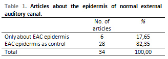
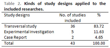
Among all articles included in this study, few were published in order to only analyze, through immunohistochemistry, the epidermis of the external auditory canal. Most of the articles which studied the normal EAC, used it just as a control sample to assess the immunoexpression of cholesteatoma antigens, either of middle or external ear (Table 1). In terms of the drawings of the studies, most included articles were transversal studies with a lower quantity of experimental researches (Table 2). In the six articles which were exclusively concerning normal EAC skin the expression of Ki-67, vimentin, PCNA, IgA, IgG, IgM and several cytokeratins were analyzed.
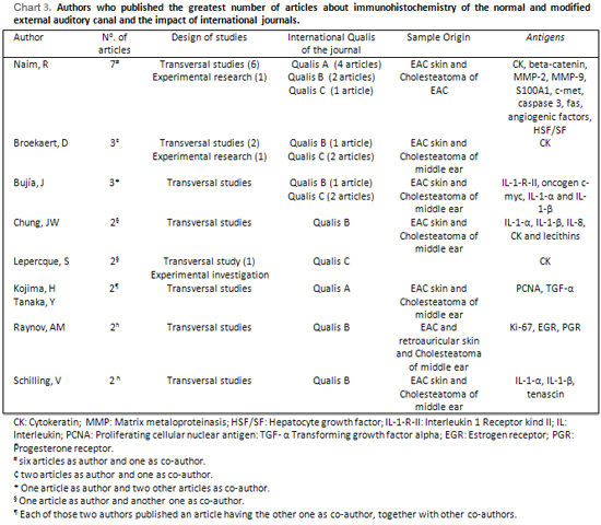
Some authors published more than one study in specific areas of this topic. The author who published the greatest number of articles was Naim, R, with seven articles about immunohistochemical analysis of cholesteatoma of external auditory canal (Chart 3).
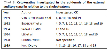
The cytokeratins were far the most investigated antigens in the included studies (Chart 4 and 5). The cytokeratins, a complex with 20 polypeptide, are intermediate filaments which make the cytoskeleton of epithelial cells. Such filaments are divided according to its molecular weight and its isoelectronic point in two subfamilies: Basic ones and acid ones. They are usually distributed in pairs in certain combinations depending on the kind of epithelium (simple, stratified or transitional), on the potential and on the relative state of keratinocyte differentiation, on the proliferative state, as well as the environmental growth conditions. For example, CK 16 is found in tissues which are under proliferation state, like in cicatricial areas, in the normal epidermis in zones which are submitted to pressure and friction (foot sole, finger pulp and heel) in the epithelium which covers the pilous follicles, in benign hyperproliferative epidermal diseases such as vulgar wart, psoriasis, actinic keratosis and seborrheic dermatitis and in malign diseases such as spinalcellular carcinoma (Ribeiro, 2004).
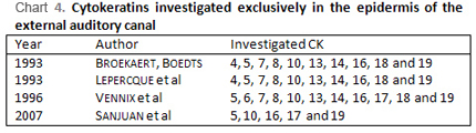
The cytokeratins, although they are the most frequently studied ones, were investigated exclusively in the epidermis of the normal external only by Broekaert, Boedts (1993), Lepercque et al (1993), Vennix et al (1996) and Sanjuan et al (2007), different from other authors who investigated them in diseases of external and mi8ddle ear, using the epidermis of the canal just as control. Broekaert, Boedts (1993), Lepercque et al (1993) and Vennix et al (1996) investigated the distribution of cytokeratins in the several parts of the external auditory canal and in the external part of the tympanic membrane, even indicating in which epithelium layer the CK were expressed. As it was expected, the three authors agreed that there is no expression of CK4 and CK13 in any region of the normal EAC, once they are specific cytokeratins of non-keratinized epithelia. Expression of simple epithelia 7, 8 and 18 were not found either. The authors found expression of CK5, CK10 and CK14 in the whole canal including the tympanic membrane, and Broekaert, Boedts (1993) and Vennix et al (1996) specified the localization of the epithelial layer in which such CK were found. Thus, the CK10 was observed in all cells of suprabasal cells and the CK5 and CK14 were found in the basal layer in the whole canal and tympanic membrane. Such cytokeratins were also present in the suprabasal layers of the bone portion of the canal, of the fibrocartilaginous ring, of the flat part of the tympanic membrane and next to the malleus (Fig. 1 and Fig. 2). Just Vennix et al (1996) investigated CK6 and CK17. The CK6, which is typical of epithelia with proliferative characteristics, was only found in the bone portion of the canal, in the suprabasal layers and in some areas of the basal layer. CK17 was found just in the bone part of the canal, in cells of the basal layer. According to the three authors, the CK16, also a characteristic of epithelia in hyperproliferative state, was not observed in the cartilaginous part of the canal, and was expressed in the most medial region of the bone portion of the canal in the cells of the suprabasal layers. In the tympanic membrane, according to Vennis et al (1996), such CK was present in the regions of the ring, flat part and malleus. Leperque et al (1993) observed a gradual reduction in the intensity of expression of CK16 in centripetal direction in the membrane. That shows a proliferative capacity in the suprabasal layers of the epidermis of the regions which are close to the fibrocartilagenous ring (Fig. 3 and Fig. 4). The CK19 was found by Broekaert, Boedts (1993) and Leperque et al (1993) in the inferior region of the tympanic annulus and next to it in the bone portion of the canal and by Vennix et al (1996) just in some cell agglomerates in such region. It is interesting to observe that, in the normal skin of the rest of the body, there is no immunosuppression of cytokeratins which are characteristics of cellular hyperproliferation, such as the CK16 and CK6, except in regions which are subject to friction and pressure in the epithelium of the pillous follicles. Sanjuan et al (2007) investigated the expression of cytokeratins 5, 10, 16, 17 and 19 in the EAC normal epidermis and after epidermis culture.. The authors observed that the patterns of expression of such filaments would be kept before and after the culture, even corroborating its localizations within the epidermal layers which were observed by Lepercque et al (1993) and Vennix et al (1996).
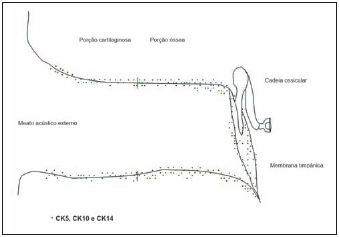
Picture 1: Distribution of CK5, CK10 and CK14 in the external auditory canal - based on articles by Broekaert, Boedts (1993), Lepercque et al (1993) and Vennix et al (1996)
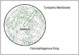
Picture 2: Distribution of CK5, CK10 and CK14 in the tympanic membrane - based on articles by Broekaert, Boedts (1993), Lepercque et al (1993) and Vennix et al (1996)
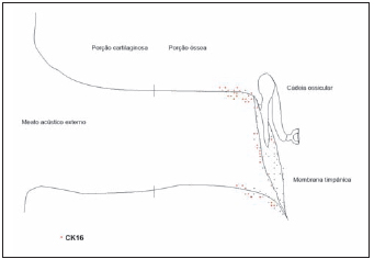
Picture 3: Distribution of CK16 in the external auditory canal - based on articles by Broekaert, Boedts (1993), Lepercque et al (1993) and Vennix et al (1996)
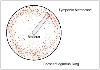
Picture 4: Distribution of CK16 in the tympanic membrane - based on articles by Broekaert, Boedts (1993), Lepercque et al (1993) and Vennix et al (1996)
Cytokeratins were also evaluated in the external auditory canal in other six studies which will be mentioned as follows, in which the authors analyze its expression in the epithelium of cholesteatoma in relation to the epithelium of the normal external auditory canal, and the latter was just a control sample. In these studies there was no specification of the regions of the canal from which the samples were taken, but most of them corroborate to the findings of Broekaert, Boedts (1993), Lepercque et al (1993) and Vennix et al (1996), who seem to have done the main studies concerning the expression and distribution of CK in the external auditory canal. Van Blitterswijk et al (1989) agree on the expression patterns of cytokeratins 10, 4, 8 and 18, and only the first one was positive in the canal. In their investigation, as well as on of Kim, Chung (1999), only CK19 presented different pattern (absent), however such fact may be explained due to the region in which the samples were obtained. According to Lepercque et al (1993) and Vennix et al (1996), the CK19 is expressed in the bone portion of the external auditory canal which is closer to the fibrocartilaginous ring, and such articles did not clarify the specification of the place where the samples were obtained. Broekaert et al (1992) also agreed concerning the positivity of cytokeratins 5, 10, 14, 16 and 19 and concerning the absence of cytokeratins 7, 8 and 18 in the EAC and tympanic membrane. Sasaky, Huang (1994) also observed the expression of CK16 in the normal external auditory canal. The samples were probably obtained of the deepest regions of the canal, once it has been well-defined that only in this region there is expression of such CK in the canal. The CK13 was not found in the epithelium of the normal external auditory canal by those authors, which is different from Lee et al (1994), who surprisingly observed the presence of cytokeratins 4 and 13 in the suprabasal layer of the epithelium of the canal. It is known that such cytokeratins are present only in non-keratinized epithelia. Such authors also found normal EAC, now as it is expected, immunosuppression of CK14 in the basal and suprabasal layers and the cytokeratins 10 and 19 only in the suprabasal layer, just like in the cholesteatoma of middle ear. Kim, Chung (1999) investigated the presence of cytokeratins in animals. In the gerbils investigated, the expression of cytokeratins 10, 13 and 16 was similar to the one observed in human samples. There was difference only in the CK4 expression, which was present, and of CK19, which was not found probably because of the sample area.
In the modified external auditory canal, the cytokeratins were investigated with positivity in cases of diseases which are common to the meatus, such as scaly cell carcinoma (unspecified CK), by Kamiya et al (1999), ceruminous adenoma (CK7), by Thompson et al (2004) and of cholesteatma of external auditory canal (CK16), by Ribeiro et al (2004).
Several antigens related to the cellular proliferation were studied in the included works. Most studies demonstrate that there is expression of such antigens in the epidermis of the normal external auditory canal. Among the authors who had the information of the place of the canal samples, some agree on the fact that such antigens, just like the cytokeratins which usually proliferate, have their expression in the EAC bone part, more precisely in the regions which are close to the fibrocartilaginous ring, with more concentration inferiorly. Before such fact, one may question whether the immunosuppression of hyperproliferation markers in the epidermis of the bone portion of the external auditory canal, next to the fibrocartilaginous ring, could be related to the differentiated ability of tissue neo-formation which occurs during the EAC embryogenesis. In addition to this, it is also questioned whether the maintenance of such capacity could have such relation with the genesis of the cholesteatoma.
Among the hyperproliferation markers, the most studied one was the Ki-67. The Ki-67 marker, a nuclear and nucleolar antigen which is closely related to cellular proliferation, was investigated in five articles about the normal external auditory canal and in four concerning the modified canal, as it will be discussed as follows. All authors found expression of such marker both in the normal and in the modified external auditory canal. Kakoi et al (1997) observed the presence of Ki-67 in the basal cells of flat and tense pars, both in the ring and in the malleus regions. On the other hand, in the epidermis of the canal, the Ki-67 was observed mainly next to the inferior ring region. Not only in the external auditory canal, but also in the tympanic membrane, the expression of such marker agree with the ones of cytokeratins 6, 16 and 19, what suggests even more the peculiar proliferative characteristic of such regions. Schilling et al (1996), Adamczyk et al (2003), Raynov et al (2005B), Hwang et al (2006) observed strong positivity for the Ki-67 in the EAC epidermis, without however specifying the localization of the samples. Such marker was also investigated in cases of canal disease by Konishi et al (2003), in one case of seborrheic keratosis, by Thompson et al (2004), in the ceruminous adenoma and by Ribeiro et al (2004), in the cholesteatoma of the external auditory canal. In such diseases, with notoriously proliferative features, there was positivity in all samples studied. Still in the EAC cholesteatoma, Sauter et al (2007) investigated the expression of Ki-67 in samples before and after the use of sulindac sulfonate and observed a reduction of the expression of such marker as the time exposure to the drug increased.
The proliferating cellular nuclear antigen, also connected to hyperproliferative states, was studied in some articles. Kakoi et al (1997), who investigated such antigen together with the Ki-67, found a similar expression pattern, having observed it mostly in the basal layers of the areas close to the fibrocartilaginous ring, including the bone portion of the canal. There is a gradual reduction of its expression in the tympanic membrane in centripetal way. Even this expression pattern is similar to the one of CK16. Kijima et al (1998), Tanaka et al (1998) and Tomita et al (2000) also found positivity of PCNA in the EAC epidermis. The first ones observed its expression in the epidermis of the canal in the basal and suprabasal layers, and the two last ones in the basal layers only. Once none of such studies had specification of the exact place from which the samples were obtained, it is not possible to affirm that only the region next to the ring has positivity to PCNA. It is expected that such antigens related to the cellular proliferation are really present in higher concentration in the regions which are closer to the fibrocartilaginous ring, both in the tympanic membrane and in the external auditory canal, once the studies makes one believe, considering the expression of Ki-67 and CK-16, that there is indeed a higher proliferative potential in the epidermis of such regions.
Other antigens related to the proliferation were studied in the epidermis of the external auditory canal, but in just one study each and without description of the sample areas. They may even more corroborate the presence of hyperproliferative capacity of the epidermis of such region. It is the case of p27 (Bayazít et al, 2001) and of PPAR-gama (Hwang et al, 2006), who were found expressed in the epidermis if the normal external auditory canal. The protein S100A1, related to hyperplasia, was also observed in the canal by Naim et al (2006).
Beta-catenin participates on the modulation of the junctions of the intercellular adhesion, whose disarrangements and reformulations are essential for the epithelial migration mechanism of the skin of the external auditory canal and for the maintenance of its integrity. Such protein is part of the system of cadherin factors of intercellular adhesion and is also directly related to the prevention of apoptosis. The beta-catenin is usually located in the zones of cellular adherence but when it is exposed to such stimuli, its translocation may occur to the nucleus. Its intranuclear dissociation allows a persistent activation of growth factors and promotes a reduction of cellular integrity. It is known that the reduction of the beta-catenin expression is closely related to the increase of tumor invasion capacity. Naim et al (2003, 2004B, 2005A) and Sauter et al (2007) were the only authors who investigated the expression of such protein. In two investigations made by Naim et al (2003, 2004B), beta-catenin was analyzed in cholesteatoma of the external auditory canal in relation to the skin of normal canal. The authors observed the immunosuppression of beta-catenin in all layers of EAC epidermis with homogeneous pattern, while in the cholesteatoma of the external auditory canal, such protein was found only in the basal layer. No expression of beta-catenin was observed in the keratin debris. Naim et al (2005A) also observed a reduction of beta-catenin reduction in cholesteatoma samples of EAC when associated to the beta transforming growth factor (TGF-
b). The co-relation between such antigen and the other previously described in this study is harmed due to the lack of specification of samples areas. However, after the work analysis, it is impossible to not observe that, besides the increase of cellular proliferation in the canal cholesteatoma, previously mentioned, there is also a reduction of the beta-catenin expression, with consequent interference in the normal process of apoptosis and intercellular adhesion. This fact may be related to the invasive characteristics of the cholesteatoma. Sauter et al (2007) investigated the expression of beta catenin in samples of EAC cholesteatoma before and after the use of sulindac sulfonate. Such authors observed not only its presence in such samples but also a reduction of its expression as the time exposure to the drug increased. Another antigen related to the cellular adhesion was also investigated.
Interleukin 1 (IL-1) is a cytokine which is found expressed in the normal epidermis and has the potential of acting as an autocrine growth factor of epithelial cells. In addition to this, it is known that such interleukin is the most powerful inductor factor of bone erosion (Schilling et al, 1992). Schilling et al (1992) and Chung, Yoon (1998) investigated the expression of two molecular species of IL-1 (IL-1-
a e IL-1-
b) obtaining positivity both in the epidermis of the canal and in the cholesteatoma epithelium. The first ones found them in all epidermis layers of the external auditory canal and of the cholesteatoma of the middle ear, with higher positivity in the cholesteatoma. The receptor II of Il-1, investigated by Bujía et al (1997) was found in the basal and suprabasal layers with homogeneous distribution both in the normal EAC epidermis and in the epithelium of the cholesteatoma, with three times higher positivity in the last one. The matrix metaloproteinasis 2 and 9, also involved in the process of bone remodeling, were found in the external auditory canal by Naim et al (2004B). One more time, in any of those articles the canal region from which the samples were taken was specified. The same thing happened with the findings of Holly et al (1995), who investigated the oncogene c-myc, Tanaka et al (1998) who investigated the expression of TGF-
a, of Tomita (2000) who investigated the p53, bax and bcl-2 of Adamczyk et al (2003), who studied the TGF-
a and the epidermal growth factor receptor (EGFR). Such antigens were found in the EAC, but just like in other articles, without specification of the exact point from which the samples were taken.
FINAL CONSIDERATIONS:After the review and the analysis proposed in this study it has been possible to conclude that:
1. There is a concentration of hyperproliferation markers in the fibrocartilaginous ring and in the regions of the external auditory canal and adjacent tympanic membrane, mainly in the inferior regions;
2. The CK16 is found in the epidermis of the normal external auditory canal just in its bone part and, in the tympanic membrane, its expression reduced in centripetal way;
3. The hyperproliferation markers Ki-67 and PCNA are also found just in the bone part of the external auditory canal, the same place of CK16 expression, and its expression reduced in the superior way;
4. Several published articles did not have their data better used and analyzed once the areas from which the samples were taken were not specified, both in the external auditory anal and in the tympanic membrane.
REFERENCES1. Broekaert D, Boedts D. The proliferative capacity of the keratinizing annular epithelium. Acta Otolaryngol. 1993,113(3):345-8.
2. Lepercque S, Broekaert D, van Cauwenberge P. Cytokeratin expression patterns in the human tympanic membrane and external ear canal. Eur Arch Otorhinolaryngol. 1993, 250(2):78-81.
3. Vennix PP, Kuijpers W, Peters TA, Tonnaer ELGM, Ramaekers FCS. Epidermal differentiation in the human external auditory meatus. Laryngoscope. 1996, 106(4):470- 5.
4. Kakoi H, Anniko M, Kinnefors A, Rask-Andersen H. Auditory epidermal cell proliferation. VII. Antigen expression of proliferating cell nuclear antigens, PCNA and Ki-67 in human tympanic membrane and external auditory canal. Acta Otolaryngol. 1997, 117(1):100-8.
5. Sanjuan M, Sabatier F, Andrac-Meyer L, Lavieille JP, Magnan J. Ear canal keratinocyte culture: clinical perspective. Otol Neurotol. 2007, 28(4):504-9.
6. Takahashi S, Nakano Y. Immunohistochemical demonstration of Langerhans cell in cholesteatoma using an antiserum against S-100 protein. Arch Otorhinolaryngol. 1989, 246(1):48-52.
7. Van Blitterswijk CA, Grote JJ, Lutgert RW, Hesseling SC, Out CJ, van Muijen GN, Fransen JA. Cytokeratin patterns of tissues related to cholesteatoma pathogenesis. Ann Otol Rhinol Laryngol. 1989, 98(8 Pt 1):635-40.
8. Broekaert D, Coucke P, Lepercque S, Ramaekers F, Van Muijen G, Boedts D, Leigh I, Lane B. Immunohistochemical analysis of the cytokeratin expression in middle ear cholesteatoma and related epithelial tissues. Ann Otol Rhinol Laryngol. 1992, 101(11):931-8.
9. Schilling V, Negri B, Bujía J, Schulz P, Kastenbauer E. Possible role of interleukin 1 alpha and interleukin 1 beta in the pathogenesis of cholesteatoma of the middle ear. Am J Otol. 1992, 13(4):350-5.
10. Sasaki H, Huang CC. Expression of cytokeratins 13 and 16 in middle ear cholesteatoma. Otolaryngol Head Neck Surg. 1994, 110(3):310-7.
11. Lee RJ, Sidey C, Narula AA, James RF. The nature of the epithelium in acquired cholesteatoma: Part 3-Cytokeratin patterns in aural epithelial and cholesteatoma cells grown in cell culture. Clin Otolaryngol Allied Sci. 1994, 19(6):516-20.
12. Schilling V, Lang S, Rasp G, Mack B, Nerlich A. Overexpression of tenascin in cholesteatoma and external auditory meatal skin compared to retroauricular epidermis. Acta Otolaryngol. 1996, 116(5):741-6.
13. Bujía J, Kim C, Ostos-Aumente P, Lopez-Villarejo J, Kastenbauer E. Enhanced epithelial proliferation due to elevated levels of interleukin-1 receptors in middle ear cholesteatomas. Eur Arch Otorhinolaryngol. 1997, 254(1):6-8.
14. Chung JW, Yoon TH. Different production of interleukin-1alpha, interleukin-1beta and interleukin-8 from cholesteatomatous and normal epithelium. Acta Otolaryngol. 1998, 118(3):386-91.
15. Kojima H, Tanaka Y, Tanaka T, Miyazaki H, Shiwa M, Kamide Y, Moriyama H. Cell proliferation and apoptosis in human middle ear cholesteatoma. Arch Otolaryngol Head Neck Surg. 1998, 124(3):261-4.
16. Tanaka Y, Shiwa M, Kojima H, Miyazaki H, Kamide Y, Moriyama H. A study on epidermal proliferation ability in cholesteatoma. Laryngoscope. 1998, 108(4 Pt 1):537-42.
17. Kim CS, Chung JW. Morphologic and biologic changes of experimentally induced cholesteatoma in Mongolian gerbils with anticytokeratin and lectin study. Am J Otol. 1999, 20(1):13-8.
18. Tomita, S. Aspectos moleculares do colesteatoma - imunoexpressão das proteínas controladoras do ciclo celular: p53, bax e bcl-2. Tese (Doutorado). São Paulo: Escola Paulista de Medicina; 2000.
19. Bayazít YA, Karakok M, Ucak R, Kanlikama M. Cyclinedependent kinase inhibitor, p27 (KIP1), is associated with cholesteatoma. Laryngoscope. 2001, 111(6):1037-41.
20. Adamczyk M, Sudhoff H, Jahnke K. Immunohistochemical investigations on external auditory canal cholesteatomas. Otol Neurotol. 2003, 24(5):705-8.
21. Naim R, Riedel F, Bran G, Hormann K. Expression of beta-catenin in external auditory canal cholesteatoma (EACC). Biofactors. 2003, 19(3-4):189-95.
22. Naim R, Sadick H, Schafer C, Hormann K. External auditory canal cholesteatoma: analysis of the integrity of the tissue structure. Int J Mol Med. 2004, 14(4):601-4.
23. Raynov AM, Moon SK, Choung YH, Hong SP, Park K. Nucleoplasm staining patterns and cell cycle-associated expression of Ki-67 in middle ear cholesteatoma. Am J Otolaryngol. 2005B, 26(5):296-301.
24. Hwang SJ, Kang HJ, Song JJ, Kang JS, Woo JS, Chae SW, et al. Up-regulation of peroxidase proliferator-activated receptor gamma in cholesteatoma. Laryngoscope. 2006, 116(1):58-61.
25. Naim R, Hormann K. The role of S100A1 in external auditory canal cholesteatoma. Oncol Rep. 2006, 16(4):671-5.
26. Kamiya M, Maehara R, Iizuka S, Yoshida T, Yamanouchi H, Yokoo H, et al. Pigmented squamous cell carcinoma with dendritic melanocyte colonization in the external auditory canal. Pathol Int. 1999, 49(10):909-12.
27. Konishi E, Nakashima Y, Manabe T, Mazaki T, Wada Y. Irritated seborrheic keratosis of the external ear canal. Pathology International. 2003, 53(9):622-6.
28. Ribeiro FAQ, Pereira CSB, Almeida R. Estudo comparativo de aspectos histológicos e imunohistoquímicos entre o colesteatoma espontâneo do meato acústico externo e o colesteatoma adquirido da orelha média. Rev Bras Otorrinolaringol. 2004, 70(5):602-7.
29. Thompson LD, Nelson BL, Barnes EL.Ceruminous adenomas: a clinicopathologic study of 41 cases with a review of the literature. Am J Surg Pathol. 2004, 28(3):308-18.
30. Naim R, Chang RC, Anders C, Sadick H, Riedel F, Bayerl C, et al.Up-regulation of beta-catenin in external auditory canal cholesteatoma. Int J Mol Med. 2005A, 15(5):801-4.
31. Sauter A, Matharu R, Braun T, Schultz J, Sadick H, Hormann K, et al. Sulindac sulfone modulates beta-catenin in human cholesteatoma cell culture. Arch Med Res. 2007, 38(4):367-71.
1. Otorhinolaryngologist doctor (Master's degree on Medicine by the Faculdade de Ciências Médicas da Santa Casa de São Paulo.
2. Doctor's Degree on Otorhinolaryngology by UNIFESP (Assistant Professor of the Otorhinolaryngology Department of the Faculdade de Ciências Médicas da Santa Casa de São Paulo.
3. Master's degree on Medicine by the Faculdade de Ciências Médicas da Santa Casa de São Paulo (Assistant Professor of the Morphology Department of Faculdade de Ciências Médicas da Santa Casa de São Paulo).
4. Master's degree on Medicine by the Faculdade de Ciências Médicas da Santa Casa de São Paulo (Head Professor of the Morphology Department of Faculdade de Ciências Médicas da Santa Casa de São Paulo).
Faculdade de Ciências Médicas da Santa Casa de São Paulo.
Mail Address:
João Daniel Caliman e Gurgel
Av. João Felipe Calmon, 1115 - Centro
Linhares-ES CEP 29900-010
Phone: (27) 3371-6836 - Fax: (27) 3329-2202
E-mail: contato@joaodaniel.com
This article was submitted to SGP (Sistema de Gestão de Publicações) of R@IO on January 31, 2008 and approved on June 3rd, 2008 at 23:12:37.