INTRODUCTIONMost of bilateral paralysis of vocal cords is due to bilateral lesion of the inferior larynx nerve. The main cause is iatrogenic lesion of the nerve during thyroidectomy or, but less frequent, during other cervical and thorax surgeries. Viral infections are less common causes of bilateral paralysis of inferior larynx nerve (1).
Bilateral paralysis of vocal cords does not normally causes damages on the vocal quality, as vocal cords lies in adduced position. Therefore, there is respiratory restriction due to a narrow glottis fold. Some of these patients need immediate surgery intervention due to acute respiratory insufficiency. Other patients can bear light to moderate dyspnea for a long period with no need of therapy.
Tracheostomy can solve dyspnea problems, by sustaining good tone and air passages protection, although it is not well seen by patients as a long term solution. Therapy procedures in these cases are challenging for the ENT professional, who needs to enlarge the respiratory area of the glottis, by preserving as much as possible phonation and sphincteric functions of the larynx.
Several surgical procedures have been suggested for treating such condition. Jackson (2), in 1922, suggested ventriculocordectomy technique through external passages. In 1946, WOODMAN (3) suggested arytenoidectomy through extralayngeal access, associated to suture of vocal process inferior horn of thyroid cartilage, by making vocal cord in lateral manner.
Endolaryngeal accesses were introduced in 1948 by Thornell (4), who proposed arytenoidectomy with Electric cautery. Arytenoidectomy and posterior cordotomy performed using CO2 laser was introduced by OSSOFF et al (5, 6) and by DENNIS AND KASHIMA (7) respectively. Endolaryngeal accesses advanced correction on bilateral paralysis of vocal cords due to its small rate of morbidity.
In 1994, RONTAL et col (8) reported a new method so-called "Laryngeal muscular tenotomy", where vocal ligament, thyroarytenoid muscle and interarytenoid muscle are sectioned by their insertions, on the arytenoid cartilage. This surgery approach aimed to eliminate adduct forces over arytenoids cartilagewith a predominance of abduct action of posterior cricoarytenoid muscle. TSUJI AND COL (9) performed such technique on 9 patients, by enlarging glottis fold and improving dyspnea on 6 patients (9).
In the current study, it will be reported a particular experience of the first author on bilateral paralysis of vocal cords therapy, by combining posterior cordotomy and partial arytenoidectomy.
MATERIAL AND METHODAfter approval by the Ethics Committee on Research, #132/4, a retrospective study of the surgical outcomes of 10 patients with lilateral paralysis of vocal cords submitted to posterior cordotomy combined with partial arytenoidectomy was performed. Surgery procedure was performed by the same professional (DHT) from 2001 to January, 2005 (Table 1). Age of patients ranged from 37 to 75, and the average was 46.5 years old. All patients presented previous history of thyroidectomy. Only one patient had been submitted to a previous tracheostomy due to acute respiratory insufficiency soon after thyroidectomy. The average paralysis period ranged from 1 to 40 years (average 11.7 year). All patients reported dyspnea at light and moderate efforts.
Choosing the side of operationPerforming nasofibroscopy before surgery is important in order to choose the side of operation. The vocal cord which occupies greater space on the glottis is preferably the operated one, i.e. the one which is more mid-placed. If both cords lie in the same position, so the one that presents any trace of mobility is chosen. If mobility of vocal cords is at the same level, doctor should then consider the ability to operate over the side which offers the best access.
Surgery ApproachSurgery is performed under general anesthesia. After tracheostomy, laryngoscope of suspension is placed with an ample exposition of the glottis. The CO
2 laser, SHARPLAN 20C model is attached to the operating microscope, which is linked to Accuspot micromanipulator, in the 3.5-watt, continuous superpulse mode.
Surgery approach is displayed in Pictures 1 to 6.
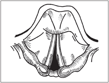
Picture 1. Endoscopic visualization of the larynx with bialteral paralysis of vocal cords in middle position.
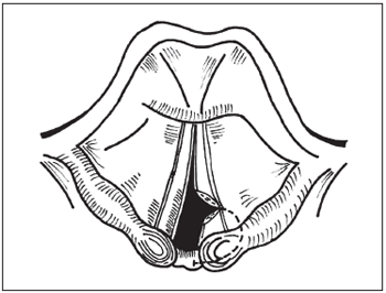
Picture 2. Posterior cordotomy.
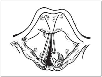
Picture 3. Submucosa disection of arytenoid cartilage.
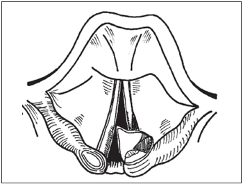
Picture 4. Vaporization of thyroarytenoid muscle fibers.
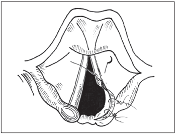
Picture 5. Bloody area covering using mucosa flap attached to fibrin adhesive and Vicryl 5.0 suture.
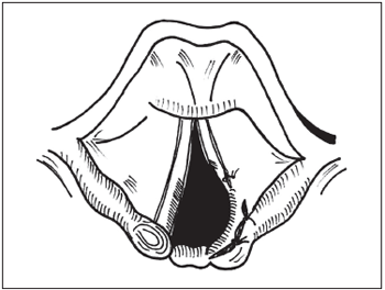
Picture 6. Final surgical aspect.
Surgery is performed under general anesthesia. After tracheostomy, laryngoscope of suspension is placed with an ample exposition of the glottis.
Surgery procedure combines posterior cordotomy (a different technique proposed by DENNIS AND KASHIMA) and subtotal arytenoidectomy, a modification of endoscopic arytenoidectomy by OSSOFF, and consists of the following steps:
Step 1: Posterior Cordotomy After exposition of glottic area, especially its posterior portion, larynx is explored with the help of palpator in order to discard fixation of arytenoids cartilage and posterior glottic stenosis (Picture 1). After protecting subglottis and trachea, posterior cordotomy is performed using a cotton piece moistened in physiological solution, by sectioning the vocal cord, before the vocal process of arytenoid, transectioning mucosa, vocal ligament and the fibers of thyroarytenoid muscle in lateral way. This section process should be lengthened in caudal manner, totally covering the elastic cone (Picture 2).
Step 2: Submucosa disection of arytenoid cartilageAn arc-way incision of mucosa is performed from vocal process area up to the interarytenoid area, by raising a caudal and medial pedicled mucous flap. Submucosa disection of the cartilage can be performed using either CO
2 laser or microscissors (Picture 3).
Step 3: Subtotal Arytenoidectomy After exposition, all vocal process is removed by using either scissors or CO2 laser vaporization. The muscular process is left intact, by keeping muscular insertions of the posterior cricoarytenoid muscles and lateral cricoarytenoid. A especial care is taken in order not to harm interarytenoid mucosa (Picture 4).
Step 4: Vaporization of the fibers of thyroidarytenoid muscle An incision measuring around 5mm of extension of the posterior and lateral part of the vocal cord, near the ventricular floor, is vaporized by using CO
2 laser in parallel to the free edge of the vocal cord, removing then mucosa and muscular tissue in submucosa way (Picture 4). Bloody edges are put together and attached with fibrin adhesive and Vycril 5.0 suture.
Step 5: Bloody area covering with mucosa flap In order to avoid stenosis and granuloma formation, the bloody area which correspond to arytenoidectomy is recovered by repositioning of mucosa flap which is attached with fibrin adhesive and Vycril 5.0 suture (using a 1.5cm needle).
All patients undergo antibiotic therapy for 10 days (cefalosporin) and corticoid therapy (hydrocortisone 500mg in the intra operative period and prednisone for 10 days in the postoperative one). After surgery, patients take prednisone and wide spectrum antibiotics for 10 days. Besides, omeprazole or pantorazole (20mg twice a day for 30 days) is prescribed in order to prevent granuloma formation.
RESULTSRestauration of air passages, by sufficient enlargement of glottic fold in order to soothe respiratory symptoms, was successful for all patients. Only one patient needed surgery due to the fact that her glottic fold became narrower 4 months later the first procedure. Such patient underwent posterior cordotomy and subtotal arytenoidectomy at the contralateral side.
Decannulation was perfromed between 4 and 8 weeks (average 5.7 weeks). 4 patients presented small granulomas. They were clinically treated with cortisone sprays and proton pump inhibitors. None of patients needed surgical revision in order to remove franulomas or cicatricial tissue.
All patients presented some loss degree of vocal quality after surgery. Although, vocal quality has not been the study aim and patients present some hoarseness and blowness, they seemed content with their tone.
DISCUSSIONSeveral surgery techniques for air passages restauration due to paralysis of vocal cords have been described in the literature.
The use of CO
2 laser together with surgical microscope represented an expressive advance when treating these patients, though it enabled a better surgery field, with no clamps competition in the interior of the laryngoscope. Besides, CO
2 laser allows a more accurate surgery, with a better control of hemostasis and less incidence of postoperative eodema and cicatricial stenosis (5-10).
Complication related to CO
2 laser use are: glanuloma formation (probably die to cellular debris left from tissue carbonization), perichondritis and perforation of endotracheal tube (11,12)
Some cares can be taken in order to reduce complication, they can be: placing cotton pieces moistened in physiological solution to protect endotracheal tube and cuffing. Those can prevent perforation of the tube and explosion risk.
Interarytenoid area should not be damaged to avoid posterior glottic stenosis formation. Recovering bloody area with mucosa flap and fibrin adhesive prevents glanuloma and cicatricial tissue formation, besides provides a quicker cicatrisation.
To some authors, performing posterior cordotomy combined with partial arytenoidectomy is a procedure which provides good ampliation of the glottic fold, satisfactory tone and low rate of symptom recurrence (12-13). Moreover, partial arytenoidectomy is associated to a better phonation and less risk of aspiration when compared to total arytenoidectomy, in which arytenoid body is removed by lowing posterior wall of larynx (13,14).
Some authors prefer uni and bilateral posterior cordotomy without performing arytenoidectomy, for being a simpler procedure, with less risk of aspiration, better vocal quality and possibility of its performance with no need of tracheostomy protection. The main advantage of it is the high incidence of re-operations, mainly due to cicatricial stenosis by leading a reduction of the respiratory area (15).
In the current study, surgery success rate, determined as improvement of respiratory symptoms and decannulation was 100%, but it went down to 90% if considered the success rate in only one procedure.
The main reason for these high rates of success is the modification introduced by the author, who not only carefully recovers the bloody area on posterior glottis with mucous flap, but also vaporizes in cotter the latero-posterior part of the sectioned vocal cord, by guaranteeing a greater glottic space. Choosing the side of operation is also another factor to be considered for the surgery success. As mentioned before, the authors of this study always operate the more mid-placed vocal fold. If both are placed in the same position, the side which presents higher trace of mobility is chosen. The latter justifies the fact that the TA muscle section minimizes its adductor tonus, enabling greater predominance of abductor tonus of the CAP muscle over arytenoid one, already mentioned by other authors (8,9).
There was symptom recurrence in one case (patient 5) 4 months after the first procedure. Patient was submitted to posterior cordotomy and partial arytenoidectomy at the contralateral side. Therapy failure was assigned to repositioning of contralateral fold for occupying greater glottic space.
Although performing tracheostomy on patients who are submitted to posterior cordotomy and partial arytenoidectomy is not well seen from the esthetic viewpoint, the authors of this study always do it. Besides considering tracheotomy enables a larger surgery field and allows visualization of posterior suture, the current authors also consider it important on postoperative period, mainly in order to avoid acute respiratory insufficiency due to occasional post operative oedema and granuloma formation.
Dysphonia grade cannot be foreseen before surgery procedure, though patients should be aware regarding vocal damages. Although dysphonia and blowness increase, current patients seemed content regarding post operative vocal quality.
Surgical complications were not reported in this study. 4 patients presented small granulomas on the surgery site, but they improved their conditions by inhaling corticoid and making use of proton pump inhibitor.
CONCLUSIONPosterior cordotomy combined with partial arytenoidectomy accounts for an efficient surgery procedure for bilateral paralysis of vocal cord therapy, by restoring (most of times and at once) air passages by hardly damaging phonatory and sphincteric larynx function.
REFERENCES1. Crumley RL. Endoscopic laser medial arytenoidectomy for airway management in bilateral laryngeal paralysis. Ann Otol Rhinol Laryngol 1993; 102:81-84.
2. Jackson C. Ventriculocordectomy, a new operation for the cure of goitrous glottic stenosis. Arch Surg 1922;4:257-74.
3. Woodman D. A modification of the extralaryngeal approach to arytenoidectomy for bilateral abductor paralysis. Arch Otolaryngol 1946;43:63-5.
4. Thornell WC. Intralaryngeal approach for arytenoidectomy in bilateral abductor vocal cord paralysis. Arch Otolaryngol 1948;47:505-508.
5. Ossoff RH et al. Endoscopy laser arytenoidectomy for the treatment of bilateral vocal cord paralysis. Laryngoscope 1984;94:1293-1297.
6. Ossoff RH et al. Endoscopy laser arytenoidectomy revisted. Ann Otol Rhinol Laryngol 1990;99:764-771.
7. Dennis DP, Kashima H. Carvon dioxide laser posterior cordotomy for treatment of bilateral vocal cord paralysis. Ann Otol Rhinol Laryngol 1989;98:930-934.
8. Rontal et al. Use of laryngeal muscular tenotomy for bilateral midline vocal cord fixation. Ann Otol Rhinol Laryngol 1990;99(10 pt 1):764-771.
9. Tsuji, DH, Sennes LU, Koishi HU, Figueredo LA. Tenotomia dos músculos adutores da laringe. Arq Otorrinolaringol 1999;3(1).
10. Khalifa MC. Simultaneous bilateral cordectomy in bilateral vocal cord paralysis. Otolaryngol Head and Neck Surg 2005;132:249-250.
11. Bizakis JG et al. The combined endoscopic CO
2 laser posterior cordectomy and total arytenoidectomy for treatment of bilateral vocal cord paralysis. Clin Otolaryngol 2004;29:51-54.
12. Ecklel HE et al. Cordectomy versus arytenoidectomy in the management of bilateral vocal cord paralysis. Ann Otol Rhinol Laryngol 1994;103:852-857.
13. Sato K, Umeno H, Nakashima T. Laser aritenoidectomy for bilateral median vocal fold fixation. Laryngoscope 2001;111(1):168-171.
14. Segas et al. Management of bilateral vocal fold paralysis: experience at University of Athens. Otolaryngol Head and Neck Surg 2001;124(1):68-71.
15. Laccourreye O. et al. CO2 laser endoscopic posterior partial transverse cordotomy for bilateral paralysis of the vocal fold. Laryngoscope 1999; 109(3):415-418.
1 Post-graduanda the Division of Clinical Otorhinolaryngology, Hospital of the Faculty of Medicine of the University of Sao Paulo.
2 Medical Otorhinolaryngologist. Medical Collaborator of Otorhinolaryngology, Division of Clinical Hospital of the Faculty of Medicine, University
of Sao Paulo.
3 PhD. Medical Assistant-Doctor of the Division of Clinical Otorhinolaryngology, Hospital of the Faculty of Medicine of the University of Sao Paulo.
4 Professor (Professor Freestyle - Teacher Discipline of Otolaryngology, Faculty of Medicine of the University of Sao Paulo).
Organization: Division of Clinical Otorhinolaryngology and the Department of Radiology of the Hospital of the Faculty of Medicine of the University of Sao Paulo.
Mailing address: Discipline of Otolaryngology, Faculty of Medicine, USP - Adriana Hachiya - Avenida Dr. Enéas de Carvalho Aguiar, 255 -- 6 th floor - Room 6021 - Sao Paulo / SP - Brazil - Fax: (11) 3865-0200 - E-mail: adrihachiya@uol.com.br
This article was submitted in Management System Publications (SGP) R@IO in the August 26, 2007. Cod. 306. Article accepted on September 24, 2007.