INTRODUCTIONBone Morphogenetic Proteins (BMPs) are members of the transforming growth factor beta (TGF-
b) superfamily with a wide range of functions in vertebrate's embriogenesis (1,2). They were first described by Urist(3), but only during the 80's techniques that permitted purification and isolation from animal bones like dogs, mice, pigs and cattle were developed (4). More recently molecular biology techniques lead to the identification of genes encoding these proteins, making possible the production of recombinant forms of various human BMPs (rhBMPs)(3,5, 6, 7).
The increasing knowledge regarding BMPs role in osseous physiology and pathology stimulated several researches about its potential clinical use. Both rhBMPS and bovine forms showed efficacy in human and animal studies, mainly in the field of orthopedics, neurosurgery, craniofacial surgery and dentistry (8,9,10,11). They have been used in a range of procedures, such as acceleration of long bone fracture repair, healing of segmental and critic defects in cranial vault, long bones and periodontal regeneration (6,9,12,13,14).
The aim of this work is to evaluate microscopically the influence of the implantation of bovine purified BMPs, using type I collagen as a carrier, in the alveolus wound healing of rats incisor tooth.
MATERIALS AND METHODS
Sample A total of 30 male albino rats (Ratus norvegicus albinus, Wistar) weighing between 180 to 190g were used in this study. The animals were divided into two groups: experimental (n=15) and control (n=15) according to the implantation or not of the BMPs.
Surgical procedures The animals were anesthetized with an intraperitoneal injection of an association of ketamine and dihidrothiazine (0,1 ml/kg and 0,05 ml/kg of body weight, respectively). The upper right incisor of all sample was extracted with special adapted instruments as described by OKAMOTO, RUSSO (15). Immediately after tooth extraction, the sockets in the experimental group were filled by bovine BMPs (Gen pro
®, Baumer SA, Brazil) using bovine type I collagen (Gen col
®, Baumer SA, Brazil) as a carrier. Both BMPs and carrier were presented as a powder that were mixed with a small volume of saline, obtaining a paste that was placed into the socket with the aid of curettes. In control group the alveolus was until it was filled by the blood clot. In both groups, immediately after this procedure, the gingival mucosa was sutured with 5-0 silk sutures, approximating the borders of the wound.
Five animals of each group were sacrificed by excessive sulfuric ether inhalation after 7, 14 and 21 days. A frontal section was made between middle and posterior thirds of the animal's head. After removing all soft tissue, the right maxilla was separated from the left by a cut along the median sagittal plane.
Laboratorial processing, microscopical and statistical analysis The right halves were cut tangentially to the distal surface of the last molar and each specimen was fixed for 48 hours in 10% formalin, decalcified in 5% formic acid, dehydrated and embedded in paraffin blocks. Longitudinal semi-serial 6¼m sections were stained with hematoxilin-eosin for histological examination. Light microscopic examination of the slices was carried out to observe the findings related to the dental alveolus healing. Thus, 5 parameters of evaluation were chosen: quantification of osteoid, immature bone, mature bone, osteocytes and osteoblasts. Each specimen was analyzed by 2 different observers. The microscopic findings received one of the following scores: 0=absence; 1=mild; 2=moderate; 3=intense. The histological examination was conducted by two blinded investigators. Data were analyzed statistically by the non-parametric U Mann-Whitney test.
RESULTS
Histological findings
Seven days - Control group The socket was partially filled by connective tissue formed by a great amount of collagen fibers , with eminent newly formed blood vessels . Intense granulation reaction was observed in some areas which was characterized by a connective tissue with intense newly formed vessels and significant number of inflammatory cells. In all specimens an intense chronic linphoplasmocytic inflammatory infiltrate could be observed. Degenerated blood clot was present in some specimens.
Seven days - Experimental group More organized connective tissue was present into the socket when compared with the control group. Granulation reaction areas were uncommon, with a chronic mononuclear inflammatory reaction ranging from mild to moderate. Newly formed blood vessels were found in all specimens. Implanted material was found as an amorphous, brown stained region in variable quantity. The connective tissue in contact or near the BMPs showed a clearly different pattern in some specimens, with condensation of the fibroblasts at these areas. In some regions, cell differentiation into osteoblasts and, consequently, deposition of osteoid matrix was identified. Trabeculae of immature bone in variable quantity could be observed in all specimens.
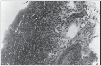
Figure 1. Control Group (Seven days) - Intense granulation reaction showing connective tissue with newly formed vessels and inflammatory cells (HE - 100x).
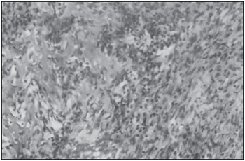
Figure 2. Experimental Group (Seven days) - Deposition of osteoid matrix indicating bone neoformation (arrow) (HE - 100x).
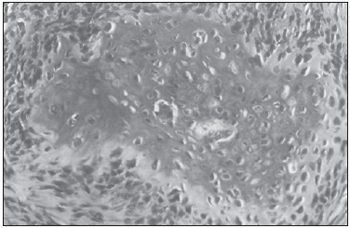
Figure 3. Control Group (Fourteen days) - Trabeculae of immature bone (HE - 200x).
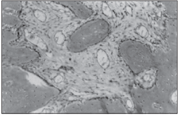
Figure 4. Experimental Group (Fourteen days) - Mature bone and fibrous connective tissue with mild quantity of newly formed vessels. (HE - 100x)
The alveolus was filled in almost all extension with well arranged connective tissue rich in collagen fibers and fibroblasts. At the medium and apical thirds of the socket was observed the presence of osteoid and immature bone with great osteoblastic activity. A mild to moderate chronic inflammatory infiltrate was present in all specimens as well as variable quantity of newly formed blood vessels.
Fourteen days - Experimental group Near total filling of the socket by immature bone was observed with great amount of active osteoblasts depositing osteoid matrix and many lacunae with osteocytes inside. In some specimens, mature bone in variable quantity could be identified. In some areas, mainly in the cervical region of the socket, there was a presence of fibrous connective tissue with mild quantity of newly formed vessels and rare mononuclear inflammatory infiltrate. Remanescents of the implanted material was observed with no apparent reaction of the organism.
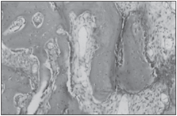
Figure 5. Control Group (Twenty-one days) - Trabecular bone filling the dental alveolus (HE-100x).
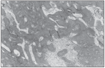
Figure 6. Experimental Group (Twenty-one days) - Dense lamellar bone observed into the socket (HE-100x).
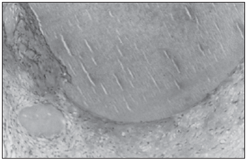
Figure 7. Experimental Group (Twenty-one days) - Remanescents of the implanted material with no inflammatory reaction (HE - 200x).
Almost all the socket was filled with immature bone but variable amount of mature bone could be seen. Significant number of active osteoblasts was found. At the cervical area of the socket the presence of fibrous connective tissue was observed with poor chronic inflammatory infiltrate and rare newly formed blood vessels.
Twenty-one days - Experimental group The socket was totally filled with mature and immature bone. In some specimens, dense lamellar bone was present with few osteoblasts in the periphery and osteocytes lacunae. It was possible to observe the implanted material predominantly in the apical region of the socket. At these areas there was no bone tissue present, being the material encapsulated by fibrous connective tissue, with the presence of mild mononuclear inflammatory infiltrate.
Statistical results The mean scores and standard deviation values of the 5 histologic parameters evaluated for alveolar osseous healing in the control and experimental groups on each observation time point (7, 14 and 21 days) are presented in Tables 1, 2 and 3, as well as the statistical differences.
DISCUSSION The alveolar healing process in rats is well documented by several studies (15, 16,17). Furthermore, this experimental animal model has been used successfully in other studies that investigated de effect of local factors on the healing of rat's dental alveolus (6,18).
The morphological events are basically the same occurring in human dental alveolus healing process, differing basically in the timing of their occurrence. The first step is the proliferation phase, when the blood clot is invaded by fibroblasts originated by mitosis of pre-existent fibroblasts and adventice cell differentiation, both present at the periodontal ligament attached to the alveolar wall. After the substitution of the clot by connective tissue, gradual substitution of this tissue by immature bone occurs, being the maturation of the bone the final phase of the process (15,16). Rat alveolar healing process takes about one third of the time of human healing process (21 days for rat dental alveolus repair against 64 days in man) (15). This healing time allows the realization of a complete study in small time period. Furthermore, this animal model has the advantage of being relatively easy to handle and to maintain and has low cost.
BMPs are extra-cellular signaling proteins and members of the transforming growth-²superfamily acting by stimulation of cell proliferation, as well as potentializing or inhibiting the response of most cells to other growth factors (6). Depending on the cellular type, BMPs actions are: inhibition or stimulation of cellular proliferation, extracellular matrix synthesis and bone formation stimulation and chemotactic cell attraction (1). BMPs osteoinductive activity is well documented in the literature. This activity was evaluated by in vitro and in vivo animal and clinical human studies, specially in long bones and craniofacial region (2,10,11,12,14,17,18,19).
There are few studies on the use of BMPs in dental alveolus. COCHRAN et al. (20) reached good results with the implantation of rhBMP-2 in post-extraction alveolus previously to implant placement in humans. However, it must be emphasized that the size sample was small and no control group was used in the study to compare the healing and osseointegration of implants in areas that did not receive BMPs. REDDI et al. (2) also used bovine BMPs in a single case report in which a trephine biopsy revealed better repair in the alveolus treated with the BMPs when compared to other alveolus that not received the proteins.
In the present study, the analysis of the 5 parameters chosen as indicative of osteoinduction showed statistically significant differences between the studied groups in the observed intervals. At 7 days period, it was possible to observe well organized bone trabeculate in the experimental group, which could not be found in the control group. The quantity of osteoid, immature bone, osteocytes and osteoblasts were significantly higher in the experimental group. Giving support to these data, our histological findings of control group are according to other studies that evaluated the normal healing process of rat alveolus (15,16). Taken together, our findings are indicatives of acceleration of the healing process due to the implanted BMPs in the experimental group.
In the 14 days period, the microscopic features of control group are compatible with the normal healing process of dental alveolus in rats at this time period. In the experimental group, more organized bone tissue was observed and the mature bone quantity was significantly higher in the experimental group showing that the implanted material was still promoting acceleration of bone repair.
At 21 days, no statistical differences between control and experimental groups were observed in the parameters related to osteoid, immature bone and osteocytes. This may be explained because at 21 days, the normal alveolar repair process is almost finished, with near total filling of the socket by bone, as described previously (15,16). However, presence of mature bone was significantly higher in the experimental group clearly showing that the healing process was in a more advanced stage.
Some comments are to be made regarding the choice of the BMPs used in this experiment. Because the concerns regarding the antigenicity and possible transmission of diseases by the bovine derived BMPs, several studies suggest that the use of recombinant human forms is preferable. However, its synthesis demands complex technology and high financial costs (5,11,21). The purified bovine BMPs have well documented osteoinductive activity even more potent than the recombinant human form (9). This osteoinductive activity is not species specific (6). The technology to obtain purified bovine BMPs is easier and cheaper than the correspondent recombinant form. Chemical processing, sterilization and the size of the proteins reduces its antigenicity and the risks of transmission of diseases (13).
The use of an appropriate material acting as a carrier to promote gradual release of the BMPs into the implantation site is a crucial point since these proteins are readily solubilized and eliminated from the tissues after implantation (4,22,23,24,25,26). Many synthetic and natural materials have been used with this aim, including deminerilized bone matrix, collagen and various synthetic polymers. Type I collagen is the main component of bone organic matrix and also the most studied carrier material both in vitro as well as in vivo. Although of xenogenic origin, purification processing of type I collagen and its associated low molecular weight lessens its antigenicity. Type I collagen is also readily resorbed by the organism. The release of BMPs by the collagen carrier is very slow, with a half-life time of 3 to 5 days (4,13). In our work, Type I bovine collagen presented as granules was used. When mixed to BMPs with saline, an easy to handle paste was obtained which was easily inserted into the socket. The carrier was efficient in releasing BMPs at the implantation site with minimal inflammatory response in the examined specimens. Although in small quantities, the presence of the material at later observation times may indicate a delay in the carrier's resorption which can be unfavorable to the later stages of bone healing process. The material was encapsulated by a fibrous connective tissue with no evidence of foreign reaction. Other studies should be performed to observe if in later periods of healing the material could be resorbed and replaced by bone.
CONCLUSION Under the conditions of this experiment, our results clearly showed that the purified bovine BMPs, using type I collagen as a carrier, presented osteoinductive activity when implanted into the dental alveolus of rats after incisor tooth extraction, accelerating the osseous healing time. These findings are encouraging regarding the future clinical use of this BMPs into alveolus after extraction, accelerating healing period and allowing the placement of osseointegrated implants in a small period of time.
ACKNOWLEDGEMENTSThis research was supported by the National Council for Scientific and Technological Development (CNPq) and Foundation for Improvement of Post-Graduation Staff (CAPES) (Brazilian Government). Also, we would like to acknowledge the help and advice of Dr. Eduardo Oliveira (PhD), Federal University of Paraíba/Brazil.
REFERENCES 1. Matzuk MM. Functional analysis of mammalian members of the Transforming Growth Factor-²superfamily. Trends Endocrinol Metabol 1995, 6:120-127.
2. Reddi A.H. Bone Morphogenetic Proteins: an unconventional approach to isolation of first mammalian morphogens. CytokGrowth Factor Rev 1997, 8:11-20.
3. Urist M. Bone formation by autoinduction. Science 1965, 150:893-899.
4. Li RH, Wozney JM. Delivering on the promise of bone morphogenetic proteins. Trends Biotechnol 2001,19: 255-265.
5. Hanamura H., Higuchi Y., Nakagawa M. et al. Solubilized bone morphogenetic protein (BMP) from mouse osteossarcoma and rat demineralized bone matrix. Clin Orthop 1980, 148:281-290.
6. Queiroz SBF, Amorim RFB, Costa ALL, Carvalho RA, Freitas RA. Proteínas morfogenéticas ósseas (BMPs): conceitos, funções e aplicações clínicas. Rev Paul Odontol 2004, 26:16-19.
7. Winn S.H, Uludag H, Hollinger JO. Sustained release emphasizing recombinant human bone morphogenetic protein-2. Adv Drug Deliv Rev1988, 31:303-318.
8. Arnaud E, De Pollak C, Meunier A et al. Osteogenesis with coral is increased by BMP and BMC in a rat cranioplasty. Biomaterials 1999, 20:1909-1918.
9. Bessho K, Kusumoto K, Fujimura K et al. Comparison of recombinant and purified human bone morphogenetic protein. Br J Oral Maxillofac Surg 1999, 3:2-5.
10. Boyne PJ. Animal studies of the application of rhBMP- 2 in maxillofacial reconstruction. Bone 1996, 19:83-92.
11. Breitbart AS, Staffenberg DA, Thorne CH et al. Tricalcium phosphate and Osteogenin: a bioactive onlay bone graft substitute. Plast Rec Surg 1995, 96:699-708.
12. Groeneveld EH, van den Bergh JP, Holzmann P et al. (1999). Histomorphometrical analysis of bone formed in human maxillary sinus floor elevations grafted with OP-1 device, demineralizaed bone matrix or autogenous bone. Comparison with non-grafted sites in a series of case reports. Clin Oral Impl Res 1999, 10:499-509.
13. Kirker-Head CA. Potential applications and delivery strategies for bone morphogenetic proteins. Adv Drug Deliv Rev 2000, 43: 65-92.] 14. Moghadam HG, Urist MR, Sandor GK et al. (2001). Successful mandibular reconstruction using a BMP bioimplant. J Craniofac Surg 2001, 12:119-127.
15. Okamoto T, Russo MC. Wound healing following tooth extraction. Histochemical study in rats. Rev Fac Odontol Araçatuba 1973, 2:153-164.
16. Ohhata H. The healing process of tooth extraction wounds observed by scanning electron microscopy and xray microanalysis. J Tokyo D College Society 1984, 84:621- 652.
17. Sakou T. Bone morphogenetic proteins: from basic studies to clinical approaches. Bone 1998, 22:591-603.
18. Heckman JD, Ehler W, Brooks BP et al. Bone Morphogenetic Protein but not Transforming Growth Factor-² enhances bone formation in canine diaphyseal nonunions implanted with a biodegradable composite polymer. J Bone Joint Surg Am 1999, 81:1717-1729.
19. Blokhuis TJ, den Boer FC, Bramer JA et al Biomechanical and histological aspects of fracture healing stimulated with osteogenic protein-1. Biomaterials 1999, 22:725-730.
20. Cochran DL, Jones AA, Lilly LC et al.Evaluation of recombinant human bone morphogenetic protein -2 in oral applications including the use of endosseous implants: 3-year results of a pilot study in humans. J Periodontol, 2000, 71:1241-1257.
21. Barboza EP, Duarte ME, Geolás L et al.Ridge augmentation following implantation of recombinant human bone morphogenetic protein-2 in the dog. J Periodontol 2001, 71: 488-496.
22. Gao T, Aro HT, Ylänen H et al. Silica-based bioactive glasses modulate expression of bone morphogenetic protein-2 mRNA in Saos-2 osteoblasts in vitro. Biomaterials 2001, 22: 1475-1483.
23. Kellomäki M, Niiranen H, Puumanen K et al. Bioabsorbable scaffolds for guided bone regeneration and generation. Biomaterials 2000, 21:2495-2505.
24. Lee JY, Musgrave D, Pelinkovic D et al. Effect of bone morphogenetic protein-2-expressing muscle-derived cells on healing of critical-sized defects in mice. J. Bone Joint Surg Am 2001, 83:1032-1039.
25. Niederwanger M, Urist M. Demineralized bone matrix supplied by bone banks for a carrier of recombinat human bone morphogenetic protein (rhBMP-2): A substitute for autogenic bone grafts. J Oral Implantol 1996 12:210-215.
26. Weber M, Steinert A, Jork A et al. Formation of cartilage matrix proteins by BMP-transfected murine. Biomaterials 2002, 23:2003-2013.
1. Masters in Oral Pathology (Professor of Anatomy of the Head and Neck, Course of Dentistry, FCRS / EC)
2. Doctor of Oral Pathology. Professor Course of Dentistry, PUC / DF.
3. PhD in Oral Pathology. Professor of the Postgraduate Program emPatologia Oral / UFRN.
4. PhD. Professor of the Postgraduate Program in Oral Pathology / UFRN.
Institution: Federal University of Rio Grande do Norte - the Postgraduate Program in Oral Pathology.
Sormani Benedict Fernandes de Queiroz
Mailing address: Avenida Santos Dumont, 6792 Papicu - Fortaleza / CE - CEP 60180-800 - Phones: (85)
3264-0025 / (85) 3265-5556 - E-mail: sormaniqueiroz@hotmail.com
National Council for Scientific and Technological Development (CNPq) and the Coordination of Improvement of Higher Education Personnel.
This article was submitted in SGP (Management System Publications) R@IO in the May 4, 2007. Cod. 248. Article accepted on June 9, 2007