INTRODUCTIONChiari malformation (MAC) belongs to a group of anomalies comprising the structures of cranial-cerebellar-medullary junction. It was first described by Cleland in 1883 (1) and, subsequently, by Chiari in 1891 (2), the latter of them had initially recognized three types: I, II, III and, later on, IV. Types II, III and IV are usually identifiable before or during birth and they can be lethal (2).
Type I (MAC I) is characterized by the descent of brain tonsils and the median portion of the inferior lobe of the cerebellum through the cervical spinal canal, below the plane of the foramen magnum In type II, cerebellar tonsils and cerebellar vermis are found to be dislocated, as well as a deformation of a part of the fourth ventricle and the medulla oblongata towards the cervical spinal canal are observed. Types III and IV include rude cranial malformations, with a partial brain and cerebellar herniation (4).
The first symptoms of type I can occur during childhood, but in most cases they appear between 30 - 50 years of age. It is mostly found in women and associated with motor, sensorial and autonomous manifestations, such as occipital cephalea, muscular atrophy and paresthesias of the upper extremities, which are aggravated by flexion, cervical extension or cough (5). Its prevalence is hard to determine, because there are many asymptomatic cases, making the epidemiologic information scarce (3).
Disequilibrium or ataxia has been noticed on 17-43% of patients (6). However rare, vertigo and nystagmus have been described as primary symptoms of MAC I (7). The otorhinolaryngological literature has few reports about the type, when hearing loss is observed in MAC I. Many cases include neurological reviews of patients showing neurological dysfunctions (8).
When diagnosing, in addition to a detailed anamnesis and a physical exam, the audiological evaluation, vestibular evidences and gadolinium-containing contrast agent for magnetic resonance (7).
The surgical treatment is regarded for patients showing progressive and debilitating symptoms, such as an increase in the intracranial pressure or an autonomic neuropathy (7).
The authors report a case showing symptoms of tinnitus, hearing loss and occipital headache, as a result of Chiari I malformation.
CASE REPORT66-year-old female patient born in the city of Belo Horizonte / MG appeared at a consultation complaining about sudden vertigo associated with nauseas and vomits, having a sensation of backward downfall and cloudy vision for two days. She also mentioned occipital cephalic, paresthesias of upper members and bilateral ear fullness without showing an improvement after taking dipyrone and acetaminophen-based pain relievers. The otoscopy, rhinoscopy and oroscopy showed no alterations. Due to the intensity of the symptoms, the patient was submitted to serotherapy and supplementary exams.
A survey was performed to disregard systemic diseases and the patient showed regular kidney and liver functions, as well as glycaemia, blood count, evidence of thyroid functions, calcium, VDRL, vitamin B12 and folic acid.
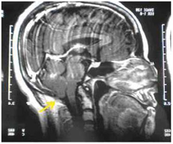
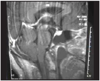
The computed tomography of the encephalic segment showed a slight reduction in the encephalic volume, and the nuclear magnetic resonance (NMR) presented a small projection of the brain amygdala through the foramen magnum, making MAC I evident (Figures 1 and 2).
The neurological evaluation confirmed the diagnosis of MAC I and it maintained a conservative treatment, because of the patient's age and the favourable evolution of the case with symptomatic drugs and vestibular rehabilitation.
After leaving the hospital, the ophthalmological evaluation including the computed visual field appeared to be within the regular limits bilaterally.
On the fifteenth day of evolution, pure-tone air and bone conduction threshold audiometry showed tone air and bone conduction thresholds ranging between 25 dB and 60 dB (250 Hz to 8000Hz), with a vocal monosyllable distinction of 96% in both ears and 35 dBNA SRT. The immitance audiometry found A-type curve with a contralateral stapedius reflex. In vector electronystamograhy, neither spontaneous nystagmus with open and closed eyes nor positional nystagmus was observed. The pendular tracking was type III, optokinetic nystagmus and vestibular normoreflex at the caloric testing review.

Figure 1. Encephalic NMR showing MAC-I.

Figure 2. Encephalic NMR showing MAC-I.
Many theories have been accepted to explain the VIII nerve involvement with MAC I. Explanations about the inner ear impairment are derived from the compression of the intracerebral cochlear nucleus and the cochlear ischemia, as well as the vestibular nerve, as a result of the torsion of the inferior portion of the brain artery or one of its branches (4). The episodes of instability, vertigo and ear fullness take us to Meniere's Syndrome, as well as the presence of any associated neurological signal or symptom (in this patient's case, paresthesias), made us think of Chiari.
Disequilibrium or ataxia has been found in 17-43% of patients (6). However rare, vertigo and nystagmus have been described as primary symptoms of MAC I (7). The otorhinolaryngological literature has few reports about the type of hearing loss found in MAC I. Many cases comprise neurosurgical reviews of patients with significant neurological dysfunctions (8).
For a diagnosis, in addition to a detailed anamnesis and physical exam, the audiological evaluation, vestibular evidences, and gadolinium-containing contrast agents for magnetic resonance are indicated. The surgical treatment is considered for patients showing progressive and debilitating symptoms, such as intracranial pressure or autonomic neuropathy.
NMR is an important supplementary diagnostic exam, in cases of suspicious MAC I, due to its several clinical similarities with a number of affections.
The diagnostic hypothesis of Chiari I must be based on complaints and clinical and image exams.
CONCLUSIONPresentation of this clinical case is a result of the fact that a rare entity was diagnosed in the patient's age group.
REFERENCES1. Cleland J. Contribution to the study of spina bífida, encefa-locele, and anencephalus. Anat Physiol. 1883, 17:257-292.
2. Chiari H. Uber Veranderungen des Kleinhirns infolge von Hydrocephalie des Grosshirns. Dtsch Med Wschr. 1891, 17:1172-1175.
3. Fonseca S. et al. Anestesia para Cesariana em doente com Malformacao Arnold Chiari tipo I e Seringomielia. Rev SPA. 2006, 16(2):24-28
4. Gonçalves da Silva JA, et al. Malformaçoes occipito-cervicais, impressao basilar, malformacao de Chiari, seringomielia, platibasia. Recife: Editora Universitária; 2003, 169-300.
5. Sperling NM, Franco RA, Milhorat T and J R. Otologic Manifestation of chiari I malformation. The American Journal of Otology. 1994, 15:0 634-638.
6. Chiari H. Ueber Veranderunger des Kleinhirns infogle von Hydrocephalie des Grosshirns. Dtsch Med Wochenschr. 1891, 17:1172-5.
7. Albers FW, Ingels KJ. Otoneurological manifestation in Chiari-I malformation. J Laryngol Otol. 1993, 107(5):441-3.
8. Rydell RE, Pulec JL. Arnold Chiari malformation. Arch Otolaryngol. 1971, 94:8-12.
1) Master in Health Sciences. Clinical Diretor of Otomed and Predecessor of the 11th and 12th-term students at Unifenas College / BH.
2) Medical Student of Barbacena Medical School.
Institution: OTOMED Clinics and Barbacena Medical School. Belo Horizonte / MG - Brazil. Mailing address: Renata Cristina Cordeiro Diniz Oliveira - Rua Ascanio Bulamarque, 199 - Bairro Mangabeiras - Belo Horizonte / MG - Brazil - ZIP Code: 30315-030 - Telephone: (+55 31) 3221-3630 - Email: dinizrenata@yahoo.com.br
Article received on November 13, 2008. Article approved on April 28, 2011.