INTRODUCTIONHearing loss is the most common findings in cases of ear abnormalities. The type and grade of this loss is related to the place of the abnormality: external, middle or inner ear.
Abnormalities on external ear are associated to the abnormalities on middle ear, as they have the same embryological origin. Though, abnormalities on inner ear are together with the ones on external ear in 15 to 20% of the cases. This can be explained by the fact that the inner ear develops itself separately and in gestational period before other ears´ development (2).
It is important to classify hearing loss as early as possible in order to determine the procedures to be taken. The possible treatment to these abnormalities involves the adaptation of hearing aids by air or bone conduction; adaptation of implanted hearing aids (integrated bone implant) or surgical rebuilding of the ear,
The surgical rebuilding of the abnormal ear is possible when there is enough development of the middle ear and when facial nerve does not recover the oval window (3). Yet, authors also report that amplification is necessary even after surgical rebuilding, as normal hearing is hardly achieved.
For bilateral abnormalities, the worry is related to speech and language development. According to Fetterman and Kuxford (4), as surgery is not recommended for children under 6, such children with this type of complication should receive suitable adaptations as early as possible in order to assist hearing stimulation needed to the development of speech and language.
Hence cases of unilateral abnormality, contralateral hearing will, if it is normal, make this development possible (4), surgery is delayed till the moment patients are able to understand the risks for their hearing and facial functions, in a way that they can decide for it by themselves.
The individuals who will not undergo surgery will be able to test hearing aids by air and bone conduction. If external auditory canal is open and big enough, a hearing aid by air or bone conduction will be adapted. Silveira et al. (6) reported the importance of investigating anatomical conditions of the external ear with abnormality, in order to verify the possibility of introducing the ear mould and the support of retroauricular hearing aid by air conduction.
Hearing aids by bone conduction are recommended for the cases in which the individual does not have anatomical conditions for air conduction adaptation. They consist of an electromagnetic vibrator compressed against mastoid, supported by an arch around the head (7).
Thus, sound wave captured by microphone is converted into electormagnetic vibrations which are send to cochlea (4).
TARGETThis study has the purpose of establishing hearing loss observed in different types of abnormalities; relating findings of CT from the temporal bone, and presenting the recommended procedure.
METHODThis study was performed at Centro de Distúrbios da Audição, Linguagem e Visão do Hospital de Reabilitação de Anomalias Craniofaciais da Universidade de São Paulo (CEDALVI, HRAC-USP), Bauru-SP (Hearing, Language and Sight Disorder Center). It was approved by the Ethics and Research Committee, protocol number 41/2003-UEP-PC.
The medical register from the individuals with abnormal ear were examined with the purpose of selecting the ones who had ENT evaluation (including CT and its result from temporal bone) and audiological evalution (threshold tonal audiometry and logoaudiometry performed at audiometer Ad-28, Interacoustic, phones TDH-39). 37 reports were selected from all these criteria together. 19 from those were children and 18 were adults.
The CT from temporal bone from individuals with abnormal ear is not part of the clinical procedure of this Center, though it is required according to the need from the ENT doctor in order to clear the implicated place and abnormal structures. As individuals come from different places, this procedure can be performed locally.
Data from ENT evaluation, from temporal bone CT and from audiological test were collected, analyzed and compared as for type of abnormality.
RESULTSFrom the 37 individuals examines, 22 were male and 15 were female. Bilateral abnormality was observed in 19 ears, while 18 presented unilateral abnormality, summing up 56 abnormal ears.
In relation to unilateral abnormalities, 13 individuals had abnormality in the right ear and 5 in the left one.
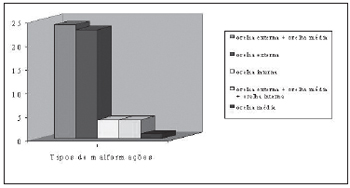
Different types of abnormalities were observed through ENT evaluation and temporal bone CT. Data related to abnormalities can be seen on Chart 1.

Chart 1. Types of malformations found in the 56 appraised ears.
External abnormality was found in 23 ears with different irregularities (Table 1).
In relation to type and grade of hearing loss observed from external ear abnormality (Table 1), only on ears with stenosis of external auditory canal had mild conductive hearing loss. The other ears (21) had moderate conductive hearing loss. From those, 1 ear presented microtia and stenosis of external auditory canal, 7 presented acoustic and external auditory canal agenesia and 13 presented microtia and external auditory canal agenesia.
Table 2 and 3 present irregularities observed from other types of abnormalities related to type and grade of hearing loss.
Procedures for the 18 individuals with unilateral abnormalities can be seen at Table 4.
Procedures for the 19 individuals with bilateral abnormalities can be seen at Table 5.
DISCUSSIONFrom the studied samples (23 individuals), we could observe a predominance of abnormality in men for unknown reasons (4,8).
According to Schuknecht (9) and Fetterman and Luxford (4), unilateral abnormality occurs from 70% to 85% of the cases, though its predominance was not observed in this study.
As for unilateral abnormality (18 ears), the predominance was on right ear (13 ears). Okajima et al. (8) and Fetterman and Luxford (4) also mentioned that, for unknown reasons, right ear is more affected in relation to abnormality.
Chart 1 shows types of abnormalities found in 56 examined individuals. These data were taken through ENT evaluation and CT, as the involvement extension can only be established by the latter. CT has become extremely important to evaluate the development of the middle ear and ossicular chain, as well as abnormalities from inner ear. This procedure helps in determining the development extension of the tympanic bone, mastoid pneumatization, mastoid, presence of oval window and stapes platinum, inner ear anatomy and surgical recommendation (5,10).
Thus, Chart 1 shows the predominance of the abnormalities from external ear (23 ears) and from external ear associated to the middle one (24 ears). External ear abnormalities are normally associated to middle ear, as they have the same embryological origin (1).
Regarding to what was mentioned above, it is possible to find different abnormalities described by the ENT doctor according to CT result. Though, after performing the audilogical evaluation, we could observe that they presented conductive hearing loss with a predominance of moderate grade (Table 1 and 2). The examined records did not present detailed descriptions regarding middle ear abnormalities.
Only 1 ear presented ossicular chain abnormality. First, the subject was submitted to an audiological evaluation, having moderate conductive hearing loss as a result. As anamnesis presented significant data, the audiological diagnosis led to a supposition of abnormality, what was infered through CT whose result could confirm the integrity of external ear and abnormality of ossicular chain. Crysdale (1) reported that children with congenital hearing loss with no microtia or atresia can present late audiological diagnosis due to invisible distortion. Hearing loss, in these cases, should be a consequence of bone distortion, ankylose or stapes fixation.
As audiological findings to abnormalities of external and middle ears and external ear associated to middle one were similar, we could see the need of performing CT from temporal bone to establish the abnormality extension (middle ear involvement).
Only 4 ears presented associated abnormalities of external, middle and inner ear whose diagnosis was achieved through CT from temporal bone. We could observe mixed and sensorineural hearing loss, in these abnormalities, and its grade ranged between moderate and profound. Cochlea abnormalities and semicircular canals exist together with atresia only in 15 and 20% of the cases, what can be explained by the fact that inner ear develops in separate way and in different gestational period from the external one. While inner ear is well developed, the external and middle ones are at the beginnig of their formation (11).
We could also see, through CT, 4 inner ears with Mondini´s abnormality. In tonal threshold audiometry, we could observe moderate sensorineural hearing loss (1 ear), severe sensorineural hearing loss (1 ear) and profound sensorineural hearing loss (2 ears). We could not find, in the literature, data related to hearing loss grade which could be compared.
Table 4 shows 10 individuals with unilateral abnormality who prefered surgery treatment in order to rebuild ear or surgery to integrate bone implant. The presence of normal contralateral hearing, possibly influenced 5 individuals, who were not candidated to surgery, to remain doing audiological control, and the other 3 prefered the adaptation of hearing aid.
In the cases of the 13 individuals with bilateral abnormality (Table 5), adaptation of hearing aid eihter through air or bone conduction was frequent. In the cases of bilateral abnormality, surgery is recommended (2), though, in this study, the criteria for selletion of candidates to this procedure were not considered.
CONCLUSIONFrom the 37 medical registers from individuals with abnormal ear, it is possible to see the predominance of abnormalities of external ear and of external ear associated to middle ear. As for hearing loss, the predominant type was moderate conductive one.
Audiological evaluation was important to see how much hearing is affected, though it was not enough foretell the type and extension of the abnormality, what has enlarged the need to perform CT from temporal bone.
Hearing aid recommendation was frequent for individuals with bilateral abnormality and surgical recommendation was frequent for individuals with unilateral one.
REFERENCES1. Crysdale WS. Otorhinolaryngologic problems in patients with craniofacial anomalies. Otolaryngol Clin North Am 1981, 14(1):145-55.
2. Bento RF, Miniti A, Butugan O. Doenças congênitas do ouvido. In: Bento RF, Miniti A, Marone SAM. Tratado de Otologia. 1ª ed. São Paulo: EDUSP, Fundação Otorrinolaringologia, FAPESP; 1998, p.135-42.
3. Gates GA, Valente M. Fitting strategies for patients with conductive hearing loss. In: Valente M. Strategies for selecting and verifying hearing aid fittings. New York: Thieme Medical Publishers; 1994, p.249-66.
4. Fetterman BL, Luxford WM. The rehabilitation of conductive hearing impairment. Otolaryngol Clin North Am 1997, 30(5):783-801.
5. Crabtree JA. Congenital atresia: case selection, complications, and prevention. Otolaryngol Clin North Am 1982, 15(4):755-62.
6. Silveira TS, Castiquini EAT, Shayeb DR, Meyer ASA. Adaptação de AASI em paciente portador de agenesia de conduto auditivo externo. Pró-Fono 2003, 15(1):95-100.
7. van der Pouw KT, Snik AF, Cremers CW. Audiometric results of bilateral bone-anchored hearing aid application in patients with bilateral congenital aural atresia. Laryngoscope 1998, 108(4):548-53.
8. Okajima H, Takeichi Y, Umeda K, Baba S. Clinical analysis of 592 patients with microtia. Acta Otolaryngol Suppl 1996, 525:18-24.
9. Schuknecht HF. Congenital aural atresia. Laryngoscope 1989, 99(9):908-17.
10. Bauer GP, Wiet RJ, Zappia JJ. Congenital aural atresia. Laryngoscope 1994, 104(10):1219-24.
11. Ribeiro FAQ. Embriologia da orelha humana. In: Caldas N, Caldas Neto S, Sih T. Otologia e audiologia em pediatria. Rio de Janeiro: Revinter; 1999, p.3-7.
1. Master (Fonoaudióloga).
2. Master (Fonoaudióloga).
3. Master (Fonoaudióloga).
4. PhD (Fonoaudióloga).
Center for Hearing Disorders, language and vision of the Hospital for Rehabilitation of Craniofacial Anomalies of the University of Sao Paulo (USP-CEDALVI-HRAC), Bauru - SP.
Mailing address: Eliane Aparecida Techi Castiquini - Street Benedito Pinto Moreira, 8-81 - Garden Panorama - CEP: 17011-110 - Bauru / SP - Telephone: (14) 3234-3563 - E-mail: elianetech@ig.com.br
This article was submitted in SGP - Sistema de Gestão de Publicações (Publication Management System) of RAIO on 17/11/2005 and approved on 17/4/2006 13:16:30.