INTRODUCTIONSecondary organic lesions, often induced by phonotrauma, are nodules of the vocal folds responsible for many of the alterations on vocal folds(1).
The low rate of glottic proportion as well as tissue features predispose laryngeal spasm and nodule formation more easily in children than in adults(4,6,8,11).
Up to the current moment, the position of the vocal nodules is not precisely defined. Interpretations regarding the situation of the vocal fold are taken by subjective experience.
The target of this study is to establish a system to delimit vocal nodules through endoscopy exam, finding them through a geometric outline.
MATERIAL AND METHODVolunteers were informed and requested for the research and after having accepted they signed the Free and Clear Consent Term. This research was submitted to the Institutional Review Boards - Universidade Tuiuti - Paraná. Of. CEP-UTP nº 066/2004 (approval protocol). All subjects involved agreed on the research and its result divulgation according to Resolução 196/96 (BRASIL. Resolução MS/CNS/CNEP nº. 196/96 de 10 de outubro de 1996.
This study was based on retrospective records from the Setor de Endoscopia do Hospital Nossa Senhora das Graças - Curitiba (Endoscopic Department) from March to July 2002.
It was chosen 86 children, 71 males and 15 females, aging from 6 months to 11 years old, with dysphonia with nodules of vocal folds.
Nodules were analyzed regarding gender, dividing material in the following ages: younger than 3 years old(5), from 3 to 4 years(21), from 5 to 6(33), from 7 to 8(22) and over 9 years old(5).
The inclusion criteria were children with vocal nodules and the exclusion one was the presence of associated laryngeal lesions.
The equipment used lists the following pieces: Mashida, ENT30TIII; a 70º laryngoscopy telescope optics, Nagashima Co. (Japan), IK-M4YIA; two VHS four heads, Victor Co. - Japan (JVC), HRJ416M; Video Monitor, Sony - Amazônia Ltda (Brasil), KV-144IB; Panasonic Printing (Matsushita Eletric Industry Co. Ltd. - Japan), AG-EP-P; stroboscopic light force 200W, Bruel-Kjael Co. Denmark, 4914; Character generator - Victor Company - Japan (JVC), CG-V60U; audio amplifier, Cygnus (Cygnbus do Brasil), AC-200. Patients were submitted to inhaling and topic anesthesia on the with lidocaine 2%.";
Regarding the photos, considering abducted vocal folds with the help of the digital pakimeter Digimatic - 500, Mitutoyo, when connected to corresponding points to anterior commissure (A) and vocal processes (BC), it was obtained a picture of a isosceles triangle (ABC) according to Pictures 1 and 2.
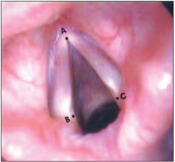
Picture 1. Normal vocal folds - A - Anterior commissure. B and C - vocal processes. AB and AC - inter-membrane space or phonatory portion of the vocal fold.
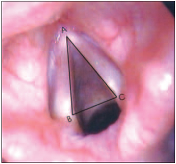
Picture 2. Normal vocal folds with delimitation of phnatory portion (Isoceles Triangle ABC). By dividing the sides AB and AC in three parts equally and joining the corresponding points to those divisions by line segments, it is possible to obtain: anterior third (AB2C2), middle third (B1B2C2C1) and posterior third (BB1C1C) of the isosceles triangle ABC, according to picture 3.

Picture 3. Anterior, middle and posterior thirds of the phonatory portion of the vocal folds. AB2C2 - Anterior third. B1B2C2C1 - middle third. BB1C1C - posterior third. By dividing middle third (B1B2C2C1) through line segment B'C', it obtains anterior-posterior thirds (B'B2C2C') and middle-posterior (B1B'C'C1), according to picture 4.
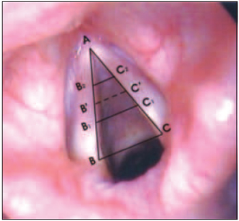
Picture 4. Middle-anterior (B'B2C2C') and middle-posterior thirds of the middle third (B1B2C2C1) of the phonatory portion of the vocal folds. B'B2C2C'- middle-anterior third. B1B'C'C1 - middle-posterior third.
The results of this study are shown in charts and tables.
Chart 1 presents percentages regarding localization of nodules on vocal folds, where it is possible to assure that such location was more frequent on the anterior third of the vocal fold, regardless gender.
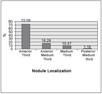
Chart 1. Sample regarding nodule localization.
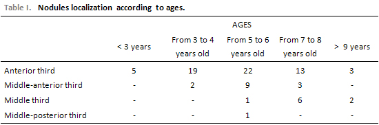
Table 1 shows that the incidence of nodule localization of the vocal folds occurred in the ages from 5 to 6.
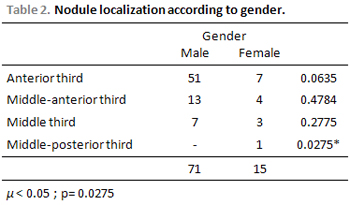
Table 2 shows data of nodule localization regarding gender with expressive result on the middle-posterior third of the vocal fold for females.
DISCUSSIONIn excised larynges one studied the relation between inter-membrane space (phonatory portion) of the glottis and inter-cartilage space (respiratory portion) of the vocal folds, confirming its variation regarding gender and age(7).
Through equivalent relation between those two parts inter-membrane space and inter-cartilage space, it was described the criteria for obtaining glottic proportion (GP)(11), from videolaryngoscopy exam images performed during comfortable inspiration in adult patients.
Parameters such as subdivision of the inter-membrane space of the vocal fold in segments which able a systematization of the anatomical localization of the nodules on the vocal fold were not established. In the current study, nodules on the anterior third of the vocal folds prevailed.
Patients of all ages presented nodules regarding the anterior third of the vocal fold, though higher incidence occurred in patients aging from 5 to 6 years, moreover, the nodules were present in all thirds of the vocal folds (Table 1).
The highest incidence of nodules on the anterior third of the vocal fold occurred in both males and females, followed by middle-anterior third and middle-posterior third in a declining sequence.
Some authors refer other places of nodules, i.e., in the transition of middle and anterior thirds of the membrane space of the vocal fold(2,3).
The presence of nodules on the anterior and middle thirds of the vocal fold was reported with no exact number of frequency in each(5,9). This study becomes original by the lack of data or information in the literature, by the used detailed parameters and by the exclusion of subjectivity regarding anatomical situation of the nodules.
The variation of the position of the nodules through this proposal answers the gaps in other studies.
This study tried to find a new systematization based on an objective analysis regarding localization of the vocal nodules, helping on locating them.
CONCLUSIONAfter analyzing and discussion over data, one might conclude that the highest incidence of nodules occurred in male patients aging from 5 to 6. Localization of nodules was more frequent in the anterior third of the vocal fold in patients aging from 5 to 6, regardless gender. The localization of nodules in the middle-posterior third of the vocal fold was expressive for females. There was higher incidence of small nodules in patients aging from 5 to 6 and from 7 to 8 both males and females. Edematous nodules were the most frequent ones especially in male patients from 5 to 6 yeas old. Bilateral nodules were the most frequent ones in males and females aging from 5 to 6 yeas old. Middle-posterior triangular gap was more frequent in patients aging from 7 to 8 for males and females. Glottic gap did not occurred in patients aging 3 years.This method of evaluation can allow its use to identify the correct position of other groups of lesion in vocal folds, such as polyps; cists, neoplasm and others.
REFERENCES1. Behlau MS, Pontes PAL. Avaliação global da voz. São Paulo, EPPM, 1990, 60p.
2. Cervantes O, Abrahão M. O nódulo vocal - conceitos atuais. RBM Otorrinolaringologia, 1995, vol 2(1): p. 12-17.
3. Colton RH, Casper JK. Compreendendo os problemas de voz: uma perspectiva fisiológica ao diagnóstico e ao tratamento. Porto Alegre: Artes Médicas, 1996.
4. Crespo AN. Coaptação glótica, proporção glótica e ângulo de abertura das pregas vocais em crianças. São Paulo, 1995. Tese de Doutorado da Universidade Federal de São Paulo - EPM.
5. Dikkers FG, Nikkels PG. Lamina propria of the mucosa of benign lesions of the vocal folds. The American Laryngological, Rhinological & Otological Society, 1999. Inc. v. 109 (10), p. 1684-1689.
6. Hast MH. Early development of te humanlaringeal muscle. In: Triglia JM, Nicolas R. Laryngitis aigües dyspneisantes de l'enfant. EMC (Elsevier, Paris) Otorhinolaringologie, 1997, vol. 20-645-E10, 5p.
7. Hirano M, Kuritas S, Nakashima T. Growth, development and aging of human vocal folds. In: Bless DM, Abbs JH. (EDS.) - Vocal Fold Physiology. San Diego, College-Hil, 1983, 22-43.
8. Leung AK, Cho H. Diagnosis of stridor in children. Am Fam Physician, 1999; 60(8):2289-2296.
9. Niedzielska G, Glijer E, Niedzielski A. Acoustic analysis of voice in children with nodule vocals. International Journal of Pediatric Othorinolaryngology, 2001. p. 119-122.
10. Peppard R, Bless D, Milenkovic P. Comparisof young adult singers and non singers with vocal nodules. J. Voice, 1988; vol.3:250-360.
11. Pontes P, Behlau M, Kyrillos LCR. Glottic configurations and glotic proportion: an atttempt to understand the posterior triangular glotic chink. Rev. Laryngol., 1994; 115:261-6.
1. Master Degree in Communication Disorder by UTP - PR (Adjunct Teacher and Coordenator of the Anatomy Department at Universidade Tuiuti - Paraná)
2. PhD in Medicine by UFP (ENT Doctor by ENT Service - HC-UFPR and Professor of Communication Disorder in the post-graduation program - Universidade Tuiuti - Paraná)
3. Mestre em Disturbios da Comunicação pela UTP-Pr (Professora Adjunta do Curso de Fonoaudiologia Universidade Tuiuti do Parana)
3. Master Degree in Communication Disorder by UTP- PR (Adjunct Teacher in the Phonoaudiology Program at Universidade Tuiuti - Paraná)
4. PhD in Anatomy by Universidade Federal de São Paulo-Escola Paulista de Medicina (Federal University) - Clinical doctor, Professor of Communication Disorder in the post-graduation program - Universidade Tuiuti - Paraná)
Universidade Tuitui do Paraná
Rua Padre oswaldo Gomes, 754, casa 3 - Fone/Fax: (41) 3335-1877 - Guabirotuba - Curitiba/PR - CEP: 80050-100
This article was submitted to SGP - Sistema de Gestão de Publicações (Publication Management System) from RAIO on September 12, 2006 and approved on December 19, 2006 21:46:42.