INTRODUCTIONTinnitus is a kind of endogenous sound sensation with no correspondent on the environment, which affects about 15% of American population (1, 2). In Brazil, we do not have definite statistical figures yet, but the American over incidence suggests the existence of approximately 25 millions of Brazilians with tinnitus (3, 4).
It is a symptom that can be caused by many otologic, metabolic, neurological, cardiovascular, pharmacological and psychological diseases, and moreover they can be present in the same individual at the same time (3, 4). Tinnitus presence can be a factor of large negative repercussion in one's life, what makes sleeping, concentration on everyday and professional activities and social life difficult. Many times it changes emotional balance of the patient, causing or worsening anxiety and depression. Despite of all this echoing and modern advances in the literature, tinnitus physiopathology had not been completely explained yet, what can implicate its treatment advance.
The association between tinnitus and hearing loss has already been described. According to different reports, 85% to 96% of patients with tinnitus present some degree of hearing loss (3, 5-8). Some authors consider the theory that tinnitus is a result of balanced mechanism to reduce disfunction of peripheral hearing pathway (9), what means, tinnitus is a consequence of hearing loss existence (10).
Only from 8 to 10% of the patients with tinnitus have normal hearing (11) and they cannot be explained by the same theory. In these individuals, isolated tinnitus presence means that it can be the first symptom of diseases usually diagnosed by hearing loss. Therefore, although they are rare, these patients have an interesting sample because their information can be related only to tinnitus and not to hearing loss, which follows the majority of cases.
However, in the vast literature on tinnitus in normal hearing, there is no longitudinal study evaluating these patients development at medium or long term to confirm hearing loss emergence, at least, up to the moment. So, pointing the importance of better evaluating these individual, the target of this study is to analyze temporal development of tinnitus and hearing on patients who have normal tinnitus and audiometry. If this is correct, the study on tinnitus can obtain importance in the specialty for justifying the attention to this complain from the professionals.
MATERIAL AND METHODThe current study and its free clear authorization term were approved by Research Project Analysis Committee of Clinical Hospital of University of São Paulo, under protocol number 630/04.
Sample SelectionThis study was traced as a longitudinal cohort. From all patients previously enrolled in Tinnitus Research Group of HCFMUSP from 1995 to 2003, it was selected the ones with the following inclusion criteria:
1. Tinnitus carrier of both sexes aging over 18 years;
2. Normal tonal audiometry on the first appointment (threshold ≤ 25 dB NA of 250 to 8000 Hz in both ears) is named evaluation 1.
From 68 patients initially eligible for the study, 32 were excluded either for lack of information or for not having attended on the appointment day in order to follow procedures, even after third call. So, final sample consisted of 36 patients with tinnitus and normal audiometry. 23 (63.8%) of them were female and 13 (36.2%) male. The average age at the time of evaluation 1 was 43.9 (SD standard Deviation =11,7ys) and, in calling time to final evaluation it was 48,9 (SD= 11,4ys).
ProceduresAll included patients already had their data related to tinnitus clinical features and preliminary audiometry from the time of initial appointment in the Tinnitus Research Group (evaluation 1) as part of medical and audiological protocol used routinely in the work.
After being informed about the research and signing free clear authorization term, patients were once again submitted to the following procedures (evaluation 2):
1. Preliminary tonal audiometry in frequencies from 250 to 8000 HZ, done by the same phonoaudiologist, using acoustic booth audiometer Midmate 622 of Madsen Eletronics correctly calibrated.
2. Simple questionnaire on current tinnitus clinical features that might be influenced from temporal evolution (localization, type, perception time, everyday activities interference, disturbance and symptoms all together).
3. Qualitative evaluation on tinnitus development through the simple question "What has happened to your tinnitus since you've began the follow-up?" Having as answer the alternatives: "annulled", "has improved", "steady" and "has worsen".
The average time break between both evaluations 1 and 2 was 3,6 years of age (ranging from 6 months to 8,5 year old; standard deviation of 2,3 years). In relation to hearing progress, patients were divided into 2 groups:
- NG (this group improved with normal audiometry): patients whose audiometry of evaluation 2 remained normal in all frequencies (threshold 25dBNA).
- AG (this group developed altered audiometry): patients whose tonal audiometry of evaluation 2 presented some alterations of tonal threshold (tested and retested) in, at least one, of the frequencies from 250 to 8000 Hz.
Analysis way of the resultsAfter digitalizing and checking of data consistency, it was made a descriptive analysis with calculation of frequencies to categorical variables and central (medium) tendencies and dispersion to quantitative variables.
Evaluation 1 and 2 data were analyzed without considering the type of treatment made in the period between them. As breaks between them are changeable, the interpretation of the data counted on the number of people/year in order to define temporal evolution of both symptoms.
To evaluate hearing development, it was calculated tonal threshold average of severe frequencies (205 and 500 Hz), medium frequencies (1000 and 2000 HZ) and acute frequencies (3000 and 8000 Hz) of each ear from each patient (t-paired test). The average frequencies in evaluation 1 between NG and AG were compared by t-test of independent variables.
To evaluate tinnitus of all studied patients and in each group (AG and NG), it was used t-paired test to compare non-binary features in evaluations 1 and 2 (age, localization and numerical scale). Non-binary features were compared using McNemar test and each patient was their own control. The non-dichotomous features of evaluation 1 were also compared by the U of Mann-Whitney test between NG and AG and dichotomous features by the Chi-square test revised in the search of prognostic features of hearing loss. Data were analyzed with the help of SPSS (Statistical Package for the Social Sciences), 13.0 version, using level of statistical significance of a = 0.05.
RESULTSI. In relation to hearing evolution The average of severe (250 and 500 Hz), medium (1000 and 2000Hz) and acute frequencies (3000, 4000 and 8000 Hz) in each ear, in bone and air pathways are in Tables 1 and 2.
16 patients, out of the 36 ones evaluated in the study, developed hearing loss in one frequency at least, and composed AG (group who developed with altered audiometry). The 20 remaining patients sustained their audiometry in normal condition and composed NG. Thus, the accumulated incidence of hearing loss was 44.5% and density of incidence of hearing loss was 38.6 by 1000 persons/year, what means, if 1000 people with tinnitus and normal audiometry were observed in one year, 38 would developed hearing loss.
In relation to degree, hearing loss was predominantly moderate in 11 patients (68.8% from AG or 36.6% from total of individuals). The 5 patients from AG presented hearing loss in light degree (31.2% from AG or 13.9% from total of individuals). In relation to the type of hearing loss, only one patient presented hypacusis, the other types were sensorineural one.
In relation to frequencies, one patient (6.3%) presented alteration only in severe frequencies and 8 (50%) presented alterations only in acute frequencies. Other 2 patients (12.5%) presented hearing loss in severe and acute frequencies and 2 patients presented it in medium and acute frequencies (12.5%). The three remaining ones (18.8%) presented alterations in all frequencies.
In relation to sides, hearing loss was predominantly bilateral, affecting 10 patients (62.5% of AG). The isolated implication of the right ear affected 3 patients (18.8%), and other 3 patients in left ear (18.9%). Bone and air pathways were more affected in acute frequencies and in left ear, what occurred in 33.3% of the patients observed or 75% of AG (table 3).
The comparison of the audiometries between evaluations 1 and 2 (t-paired test) showed an expressive difference between the average of acute frequencies (3000 and 8000 Hz) in both pathways, in both ears, and in medium frequencies (1000 and 2000 Hz) in bone and air pathways, indicating that the tonal threshold are, in fact, higher in evaluation 2 than in evaluation 1. In acute frequencies, the increasing average between audiometries was 3.3 dB for right ear (p=0.02) and 4.8 dB for left ear (p=0.01), as it is shown in table 4.
The comparison of averages of the tonal thresholds in evaluations 1 between NG (n=20) and AG (n=16), through t-test to independent variables, showed an expressive difference between the average of acute frequencies in both ears in bone and air pathways in AG. In the right ear, the average difference was 5.5 dB for the two pathways (CI 95%: 2,3-8,6; p=0.001) and, in the left ear, 4.9 dB (CI 95%: 1,2-8,4; p=0.01). The same event occurred with the average of medium frequencies at left, with an average difference of 3.7 dB (bone pathway) and 4.1 dB (air pathway), as it is shown in table 5.
The evolution of hearing loss occurred after 3.5 years of age, as a rule (SD = 2.7ys), between 6 months and 8.5 years of age. The free medium survival time of hearing loss was 5.6 years (SD = 0.6ys). Picture 1 represents Kaplan-Meyer curve showing the time up to the event, in this case, hearing loss.
II. In relation to tinnitus development IIA. Feature evaluations of tinnitus in evaluation 1 and 24 patients, out of the 36 included, did not have in the recorded their complete data on tinnitus from initial evaluation because the medical and audiologic protocols used were developed during the years). That is the reason why they were not excluded from the study, only from this partial analysis. Thus, from the 32 individuals analyzed, the tinnitus features from evaluation 1 were:
a) Localization: 18 (56.3%) patients presented bilateral tinnitus.
b) Type: 16 (50%) patients presented single and constant tinnitus.
c) Time of perception: 15 (46.9%) patients already presented tinnitus for more than 5 years when were admitted in Tinnitus Research Group.
d) Daily activities interference: the main sphere of interference was sleep quality (62.5%).
e) Discomfort: the punctuation of numerical scale from 0 to 10 points ranged from 3 to 10, with the average of 6.6 and SD of 2.4 points.
f) Concomitant symptoms: an expressive number of patients (40.6%) presented dizziness associated to tinnitus.
In evaluation 2, made with breaks of about 3.6 years (SD of 2.3ys) after evaluation 1, the predominant features of tinnitus from 26 patients, who answered the questionnaire, were:
a) Localization: 13 (50.0%) patients presented bilateral tinnitus, with no expressive difference in relation to evaluation 1.
b) Type: 17 (65.4%) patients presented single tinnitus, but that changed into intermittent (53.8%), with difference statistically expressive in relation to evaluation 1 (p=0.02) (McNemar test). The cases of pulsatile tinnitus increased from 12.5% in evaluation 1 to 26.9% in evaluation 2.
c) Daily activities interference: 14 patients (53.8%) reported interference when sleeping, with no expressive difference in relation to evaluation 1.
d) Discomfort: the punctuation of numerical scale ranged from 0 to 10 points, with average of 6.2 and SD of 2.8 points, with no statistically expressive difference in relation to evaluation 1.
e) Concomitant symptoms: 6 (23.1%) patients started reporting hypacusis. Therefore, despite of this complaint, 4 of them belonged to the group which sustained normal audiometry (NG).
IIB. Qualitative Evaluation on tinnitus developmentConsidering qualitative evaluation of tinnitus development (annulment, improvement, not changed or worse), 12 patients (46.1%) presented annulment or improvement of tinnitus and only 1 (3.8%) reported it worse (picture 2).
IIC. Comparison of tinnitus features between patients of NG and GAAfter understanding the audiometric result of evaluation 2 and classify the individuals in groups NG and AG, it was made a comparison of tinnitus features in the following situations:
a) Between NG and AG groups in evaluation 1 and 2 (Mann-Whitney test to age, localization and numerical scale; Chi-square to the other parameters): there was no statistically expressive difference in the average age of the patients from NG (46.3 years; SD of 11.1 years) and from AG (52.2 years; SD of 11.2 years) nor in features of evaluated tinnitus.
b) In each group in evaluation 1 and 2: in NG, there was no expressive difference of the observed parameters (t-paired test to age, localization and gravity; McNemar test to the other variables). In quantitative evaluation 1 and 2 of AG, there was an expressive difference (p=0,018) between discomfort average of tinnitus in evaluation 1 (7.2; SD=2.0) and in evaluation 2 (6.2; SD=2.8). In qualitative evaluation, 60% of the patients reported improvement of tinnitus and the others sustained unaltered.
DISCUSSIONThe Tinnitus Research Group of the Otorhinolaryngology Ambulatory of HCFMUSP (Hospital of São Paulo University) presents a kind of reference service and their patients are routinely submitted to a medical and audiologic protocol. That enables the evaluation of clinical features of tinnitus and correlate symptoms, as their repercussions in patient's life, facilitating the main diagnostic suspicions and the initial guiding of each case. In previous work, we studied clinical and epidemiological features from the 150 first patients assisted in our Group (12), independently from audiometric results. In that occasion, we observed the predominance of bilateral, single with long time of development tinnitus, what goes with our current findings concerning to patients with normal tonal audiometry.
In the last few years, we have assisted large number of patients with tinnitus and normal audiometry and we have asked ourselves how it would be the development in medium and long term of these individuals. Thus, the current study enabled the composition of descriptive profile of temporal development of hearing and tinnitus in these patients, who were admitted with normal audiometry in the first appointment period.
In our work, the predominance of patients with tinnitus with normal audiometry is only 7.4% of all assisted patients (13). This result is compatible to Barnea et al's findings (11), confirming that this association is rare.
It was not all initially enrolled patients who sustained a regular follow-up in the period between evaluations 1 and 2. Some of them were either using medication or under TRT (Tinnitus Retraining Therapy), others were already discharged from hospital for having their condition stable and others had been abandoned the work. Therefore, we chose to evaluate symptom development independently from type of treatment at which the patient had been submitted.
In evaluation 1, the majority of patient presented tinnitus complaint for over 5 years, what showed delay for specialized treatment. This information is important when analyzing that the majority of patients did not see an ENT doctor for the first time. Many had already been assisted in other services and been oriented to get accustomed to tinnitus.
In the course of time, the tinnitus features as localization, type and area of interference remain the same, though the event becomes predominantly intermittent. We cannot compare this development with literature, as we did not find longitudinal studies of patients with tinnitus and normal audiometry.
Common ENT anamnesis usually refrains from investigating the presence or absence of tinnitus, and it does not give details how much it causes discomfort or interference in patient's life. In the return appointments it is made a qualitative evaluation of symptoms (annulment, improvement, not changed or worse). Since the beginning of the Tinnitus Research Group, the medical and audiometry evaluation protocol has been adding investigation of discomfort degree by numerical scale from 0 to 10 in a systemic way. This quantitative evaluation has become a coadjuvant-interesting tool when choosing therapeutic strategy and when monitoring result development. For instance, two patients who present tinnitus improvement can reach different degrees of that development, in other words, the first can improve from 9 to 7, and the second from 9 to 3.Therefore, it can occur dissociation of obtained results by qualitative and quantitative evaluation, justifying our choice for both. In this study, there was an improvement of the qualitative evaluation in many of the individuals, though there was not a quantitative decrease of tinnitus discomfort by the numerical scale 6.6 to 6.1, t-paired test)
Qualitative evaluation pointed that, despite the fact that large number of patients developed hearing loss, tinnitus worsening was an exception. In this study, the only patient who reported tinnitus worsening did not present hearing loss. On the other hand, among the patients who developed hearing loss, the majority reported improvement of qualitative and quantitative tinnitus. Thus, we understood that there is no relation between audiometric worsening and qualitative tinnitus development, what differs from Barnea et al's findings (11).
NG and AG patients presented similar average of age, what makes hearing loss related to age almost impossible. This reinforces the importance of tinnitus as first symptom of a hearing disfunction. This was observed by McKee and Stephens (1992) (14) and Castello (1997)(15), who related tinnitus to an initial cochlear lesion by lesion on external ciliated cells, evaluated through otoacoustic emissions. As the application of this test was not a routine in the time of patient admission in the Tinnitus Research Group, we did not evaluate such results. Therefore, we agree on that otoacoustic emissions test in patients with tinnitus and normal tonal audimetry can be an early evaluation to detect cochlear alterations in relation to hearing loss appearing.
Audiometric evaluation showed that individuals who developed hearing loss (AG) already presented higher threshold in acute frequencies in initial evaluation, though following normal rate. This information can be interesting if we consider individuals with tinnitus and normal audiometry with threshold of acute frequencies closer to normality limit bear worse prognostic n relation to acquiring hearing loss.
In the period of follow-up, hearing loss was predominately moderate, bilateral and in acute frequencies. In spite of the audiometric curve can help diagnostic suspicious elaboration in some cases, descending sensorineural hearing loss is a type of finding frequent cochlear development and can describe a range of differential diagnosis.
Hearing loss follows tinnitus in the majority of cases (3, 5-8) and often causes favorable conditions to its appearing. Thus, some authors consider that tinnitus results from compensation mechanisms to disfunction of peripheral hearing pathway (9), what is, tinnitus is a consequence of the existence of hearing loss (10). On the other hand, patients with tinnitus and normal audiometry cannot be included in this theory. From our viewpoint, patients must have their hearing monitored, as tinnitus can imply the first alarm of hearing pathway disfunction, independently from therapeutic strategy chosen.
CONCLUSION Making evaluation of temporal development of tinnitus and hearing in patients with normal tonal audiometry, we concluded that:
1. Tinnitus neither showed worsening in the course of time, nor expressive alterations of its main features.
2. A considerable number of patient (44.5%) developed hearing loss in one frequency at least, and that was bilateral, moderate and in acute frequencies.
These findings show the tinnitus importance as first symptom of cochlear disfunction, before hearing loss establishment.
ACKNOWLEDGMENTSWe would like to thank Dr. Jeane Ramalho and Renata Marcondes for helping us in calling patients, and Dr. Desidério Favarato for the help on the statistical analysis.
BIBLIOGRAPHY1. NATIONAL INSTITUTES OF HEALTH. National Strategic Research Plan: Hearing and Hearing Impairment. Bethesda, U.S. Department of Health and Human Services, 1996.
2. SEIDMANN MD, JACOBSON GP. Update on tinnitus. Otolaryngol Clin North Am, 29:455-465, 1996.
3. SANCHEZ TG, FERRARI GMS. O controle do zumbido por meio de prótese auditiva: sugestões para otimização do uso. Pró-Fono Revista de Atualização Científica, 14(1): 111-118, 2002.
4. SANCHEZ TG. Zumbido: Análise crítica de uma experiência de pesquisa. São Paulo, 2003 (Tese de Livre-Docência, Faculdade de Medicina da Universidade de São Paulo).
5. FOWLER EP. Head noises in normal and in normal and disordered ears: significance, measurement, differentiation and treatment. Arch Otolaryngol, 39:498, 1944.
6. REED GF. An audiometric study of two hundred cases of subjective tinnitus. Arch Otolaryngol, 71:74-84, 1960.
7. SHEA JJ, EMMETT JR. The medical treatment of tinnitus. J Laryngol Otol Suppl, 4:130-138, 1981.
8. ANTONELLI A, BELLOTTO R, GRANDORI F. Audiologic Diagnosis of central versus eighth nerve and cochlear auditory impairment. Audiology, 26:209-226, 1987.
9. JASTREBOFF PJ. Phantom auditory perception (tinnitus): mechanisms of generation and perception. Neurosci Res, 8:221-254, 1990.
10. SANCHEZ, TG; FERRARI, GMS. O que é o zumbido? Em: Samelli, AG. Zumbido: Avaliação, Diagnóstico e Reabilitação (Abordagens Atuais). 1ª ed. São Paulo: Lovise; 2004. p.17-22
11. BARNEA G, ATTIAS J, GOLD S, SHAHAR A. Tinnitus with normal hearing sensitivity: extended high-frequency audiometry and auditory-nerve brain-stem-evoked responses. Audiology, 29:36-45, 1990.
12. SANCHEZ, TG; BENTO, RF; MINITI, A; CÂMARA, J. Zumbido: características e epidemiologia. Experiência do Hospital das Clínicas da Faculdade de Medicina da Universidade de São Paulo. Rev. Bras. Otorrinolaringol., 63(3):229-35, 1997
13. SANCHEZ TG; MEDEIROS IRT; LEVY CPD; RAMALHO JRO; BENTO RF. Zumbido em pacientes com audiometria normal: caracterização clínica e repercussões. Rev. Bras. Otorrinolaringol. No prelo 2005.
14. MCKEE GJ, STEPHENS SD. An investigation of normally hearing subjects with tinnitus. Audiology, 31(6):313-7, 1992.
15. CASTELLO E. Distortion products in normal hearing patients with tinnitus.
Boll Soc Ital Biol Sper. 1997 May-Jun;73(5-6):93-100.
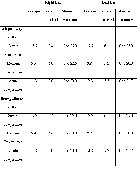
Table 1: Tonal threshold average of initial audiometry in patients admitted in Tinnitus Research Group from 1994 to 2003 with normal audiometry (n = 36)
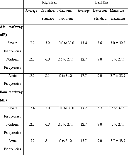
Table 2: Tonal threshold average of initial audiometry in patients admitted in Tinnitus Research Group from 1994 to 2003 with normal audiometry (n = 36)
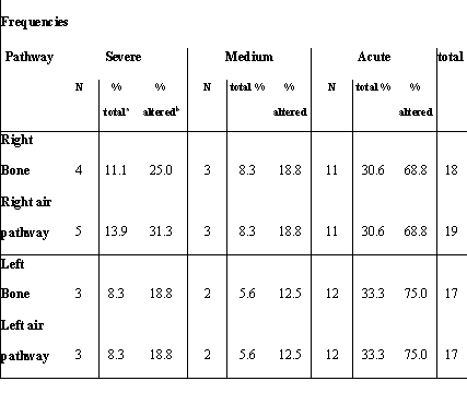
Table 3: Localization versus altered audiometric frequencies
a) % On total of patients (n=36);
b) % On patients with audiometric alteration (n=16)
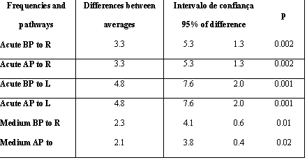
Table 4: Comparison between frequencies averages (evaluations 1 and 2)
BP: bone pathway; AP: air pathway; R: right; L: left
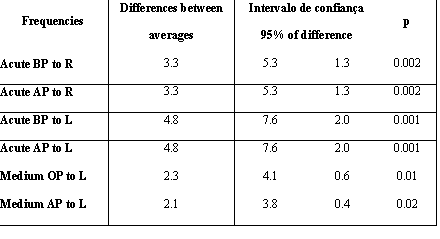
Table 5: Comparison between frequencies averages in evaluation 1 between NG (normal group) and AG (altered group)
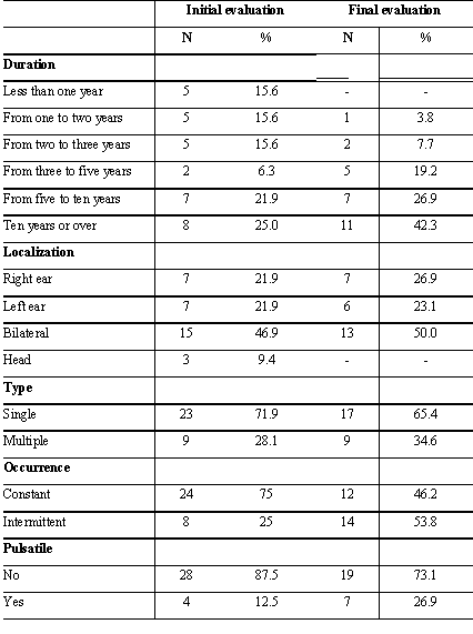
Table 6: Tinnitus clinical features of patients with normal audiometry admitted in Tinnitus Research Group from 1995 to 2003 (n=32 in initial evaluation and 26 in final evaluation).
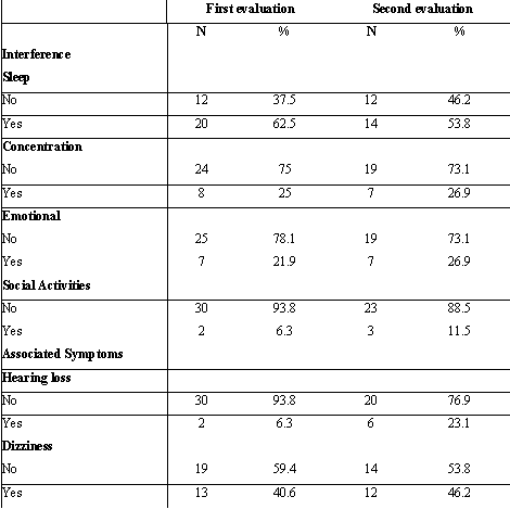
Table 7: Interference areas of tinnitus and associated symptoms of patients with normal audiometry admitted in Tinnitus Research Group from 1995 to 2003 (n=32 in initial evaluation and 26 in final evaluation).