INTRODUCTION Spontaneous pneumomediastinum is clinically rare, affecting around 1 in every 700-1200 patients in hospital(1). The main causes reported in the literature are due to intense physical activities, labour, pulmonary barotrauma, deep diving or snorkling, intense cough, cocaine inhalation, vommiting, conculsions(2,3,4). A retrospective study carried in an American Hospital showed that the main cause of pneumomediastinum was due to drug inhalation(5).
The air in the air passages can undergo pressure raise and dissect pharynx, causing cervical subcutaneous emphysema, pneumomediastinum and even pneumopericardium.
The case described reveals importance due to its rarity and its semiology. The target of this study is to describe a rare complication of the increase of intrathoraxical pressure with dissection of superficial plains having cough as a consequence and also to alarm specialists for such diagnosis on emergency cases.
CLINICAL CASE KMTC, female, 42, Caucasian, searched for ER complaining of intense odynophagia, cervicalgia and pain on interscapular area which became worse when inhaling. She had felt those for 12 hours, and all was caused by irritative dry cough which had started 3 days before.
She did not have fever, adynamia or expectoration, denied tobbaco, alcohol and drug use, chronical diseases, traumas or recent surgeries.
Physical exam presented anxiety, pains and limited cervical movements. Vital signs were stable. She presented light hyperemia of oropharinx posterior wall and light bulging on bilateral anterior-inferior cervical area which would crepitate when touched, and indirect laryngoscopy with no alterations. The cardiac auscultation revealed creptations which were syncronic to cardiac systole (Hamman Sign) and the pulmonary auscultation was normal.
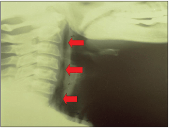
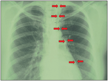
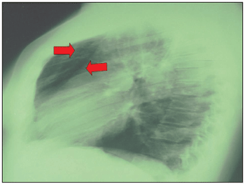
X-ray from the thorax and cervical area showed the presence of pneumomediastinum, pneumopericardium, pre-tracheal subcutaneous emphysema and presence of air on retropharyngeal space.
Patient was treated with painkillers and oxygen, remained under watching for three hours and was released from hospital after improvement of symptoms (she did not need hospitalization due to the absence of a more serious problem). She was under observation for around three hours, with relative improvement of the symptoms and good evolution. The use of oxygen was necessary, maily as hospital routine for cases of dyspnea, despite patient presented goog oxygen saturation. As she did not present either clinical, radiological signs of infection nor normothermia, no antibiotics or anti-inflammatories or any kind of medication were suggested.
Patient is under clinical observation and has not presented any new symptoms for three years.
DISCUSSION Subcutaneous emphysema associated to pneumomediastinum was first described in 1850 by Knott, in patients with cough episodes(3). Since then, theories have been suggested in order to explain its physiopathology. Marcklin suggested that intra-alveolar hypertension caused by sudden and repeated glottic closing and air passage obstruction would lead to a breakage of terminal alveolus increasing intra-thoracic pressure. Its consequence would be a flow-out of such air previously repressed in the alveolus to the interstitial pulmonary space and dissection through the vascular spaces which would lead the air to the mediastinum. Morere, in 1966, suggested that cough would unbalance capillary lung pressure. Alveolar breakage and air going into interstice, would cause dissection through vascular planes and eventually towards mediastinum. A third option would be the breakage of likely pre-existing subpleural bubbles or cysts. In general terms, therefore, there really is an increase of pressure on the alveolus by leading an air low-out(2,3).
The clinical condition is related to lung symptoms, and 82% of patients present dyspnea or thorax pain. The other symptoms pressed-throat felling; odynophagia; neck pain or dysphagia; short-term back, shoulder or neck pain and then worsened by inspiration. Around 88% of patients present subcutaneous emphysema and/or Harman sign at physical exam. Subcutaneous emphysema can extend through neck, face, tongue, axilla, arms and chest. There may be abafamento das cardiac murmurs and of precordial percussion. Hamman sign is featured by the presence of estertores or syncronic creptations with the auscultation of the cardic murmurs on the precordium(5,6).
The radiological study of cervical and thorax areas confirm the presence of air in para-pharynx and mediastinum. It is important to remember the lateral incidence of it, though 50% of patients with pneumomediastinum are not detected on anterior-posterior incidence6. Esophagus exam with barium can be used in order to avoid possible esophagus breakage as being the cause of pneumomediastinum. In a study with 36 cases, the importance of radiography was taken as the main diagnosis method(7).
Kirchner points out six important steps on pneumomediastinum diagnosis: substernal pain; subcutaneous or retroperitoneal emphysema; abafamento of cardiac murmurs; Hamman sign; mediastinum pressure increase with dyspnea, cyanosis, venous ingurgitation or blood circulation failure, and radiological evidences of air on the mediastinum(6). Despite all that, it is necessary to be suspicious to diagnose(7).
In general terms, this condition is self-limited, and conservative procedures are enough for its therapy. Pain killers and 100% oxygen are basically the procedures to be taken when necessary. Therefore, some literatures show that oxygen is not necessary. Most of time oxygen saturation is satisfactory and the use of painkillers is enough to soothe pain and muscle tension. Occasional therapy should be done(6).

Figure 1. Simple profile cervical X-ray - exam which reveals retropharyngeal emphysema.

Figure 2. Simple chest anterior-posterior X-ray - exam which reveals pneumomediastinum.

Figure 3. Simple chest profile X-ray - exam which reveals anterior pneumomediastinum.
This is an important differential diagnosis to be done at ER, as it can be wrongly taken by infection, allergic and even neoplasm processes, and patients can present sudden and intense symptoms. Despite the condition of symptoms, it is self-limited and benign, and therapy is simple and even palliative. Correct and previous diagnosis, though, can keep patient calm and it is also less expensive for hospitals.
REFERENCES1. Sleeman D, Turner R. Spontaneous pneumomedistinum with alteration in voice. J Laryngol Otol 1989; 103:1222-3.
2. Wiesner B; Frey M. Spontanes Pneumomediastinum bei Asthma bronchiale. Schweiz Rundsch Med Prax 2006;95(10):369-73.
3. Parker GS, Mosborg DA, Foley RW, Stiernberg CM. Spontaneous cervical and mediastinal emphysema. Laryngoscope 1990; 100:938-40.
4. Avaro JP, DJourno XB, Hery G, Marghli A, Doddoli C, Peloni JM, et al. Pneumomédiastin spontané du jeune adulte: Ferreira LMBM une entité clinique bénigne. Rev Mal Respir 2006;23 (1 Pt 1):79-82.
5. Chujo M, Yoshimatsu T, Kimura T, Uchida Y, Kawahara K Spontaneous pneumomediastinum. Kyobu Geka 2006;59(6):464-8.
6. Jabourian Z, McKenna EL, Feldman M. Spontaneous pneumomediastinum and subcutaneous emphysema. J Otolaryngol 1988; 17:50-3.
7. Campillo-Soto A, Coll-Salinas A, Soria-Aledo V, Blanco- Barrio A, Flores-Pastor B, Candel-Arenas M, et al. Neumomediastino espontáneo: estudio descriptivo de nuestra experiencia basada en 36 casos. Arch Bronconeumol 2005;41(9):528-31.
1. Resident ENT Doctor at Hospital Geral de Fortaleza - CE.
2. ENT Doctor - Head of ENT Resident doctor program at Hospital Geral de Fortaleza - CE.
Hospital Geral de Fortaleza - CE (General Hospital - CE - Brazil)
Lidiane Ferreira
Address: Av Washington Soares, 5353 bloco 4, apto. 202 - Fortaleza/CE - CEP: 60830-030 - Fax: (85)34866400 - E-mail: lidianembm@yahoo.com.br