INTRODUCTIONThe temporal bone malignant tumors are uncommon and occur in less than 0.2% of the head and neck neoplasms (2). The spinocellular and basocellular carcinoma are the most frequent (3) and the adenoid cystic carcinoma may rarely occur (1).
The adenoid cystic carcinoma is a malignant tumor typical to minor salivary glands; it's rare in the external auditory meatus and originate in the ceruminous glands. This tumor may also arise from lacrimal gland, bronchus, breasts, genitals and intestine. It tends to be locally invasive and of slow growth, but has a local recurrence tendency. Even with the local control of the disease, it's known to have a late metastatic presentation (6). In the past this tumor was frequently diagnosed and treated wrongly, because it had a deceiving behavior and appearance and was mistaken for a benign disease.
It equally affects men and women (1), may appear at any age, except for children, and is proven to occur normally between the fifth and the seventh decade of life (3).
The main initial symptom, in 90% of the patients, is earache (1). Other symptoms also occur, such as bleeding, otorrhea, dizziness, deafness and facial paralysis (3). The tumor mass may present as a polyp, ulceration, granulation tissue or simply as a discreet subepithelial elevation. The regional metastases are mainly for subdigastric lymph nodes, uncommon remotely, and, when occurring, the most affected location is the lung (1).
To obtain the diagnosis computed tomography is carried out and it is confirmed upon histopathological exam (2).
The treatment is essentially surgical, combined or not with postoperative radiotherapy (4).
LITERATURE REVIEWThe adenoid cystic carcinoma was first described in 1859 by Billroth, who called it cylindroma. In 1894, Haug used for the first time the term adenoid cystic carcinoma to identify this tumor, and this denomination was restated in 1942, by Spies, Quattlebaum, Foote and Frazell. The external auditory meatus tumors classification was formulated by Wetli e col. in 1972. The classification was based on aspects found in the electronic microscopy, biological activity and the answer to several modalities of instituted treatment. Four types of standards arose from this research, including: ceruminous adenoma; ceruminous adenocarcinoma; adenoid cystic carcinoma and pleomorphic adenoma (1).
Type I or Ceruminous Adenoma - benign, localized, of well-differentiated ceruminous glands, cystic or papillary. It doesn't invade adjacent tissues, but, when not fully excised, frequently presents recurrence (4).
Type II or Ceruminous Adenocarcinoma - has the same histological presentation as the previous one, in some cases shows pleomorphism and mitotic activity, infiltrative nature and involves soft tissue and bone with a high incidence of recurrence after partial surgical removal. It may provoke intracranial or remote metastases (4).
Type III or Adenoid cystic carcinoma - is the most common type, of higher malignancy, and the only one proven to produce remote metastasis. According to Ducheteau and cols. (1976), pulmonary, renal, cutaneous and osseous metastases are common. Histologically, it presents a malignant aspect with niches of small dark cells, inside which there is cystic space or hyaline material. It presents a large tendency to invade nervous structures (4). May be categorized into three histological subtypes based on the growth pattern: tubular, cribriform and solid (5).
Type IV or Pleomorphic Adenoma or Mixed Tumor - is more uncommon, lobulated, well-delimited, presents strings and nests of epithelial cells surrounded by myxoid and/or pseudo-cartilaginous stroma of myoepithelial, benign origin, but may also recur with multiple nodes where not fully removed (4).
Intracranial and remote metastases occur generally with a higher frequency than lymphatic metastases, which are uncommon (4).
Shotton et al assume the cranial base may be invaded through three ways: Eustachian tube (peritubal space), maxillary nerves and internal carotid artery (6).
Surgery remains as the adenoid cystic carcinoma treatment, with a growing interest in the technique of sentinel lymph node biopsy (7). When the tumor is located exclusively in the external auditory meatus, the treatment without bone destruction is block resection with removal of the osseous meatus with the cartilages, modified radical mastoidectomy of malleus and incus, parotid, the entire muscular, lymphatic contingent and nervous structures with facial preservation followed of irradiation. In the major tumors, a more radical resection is required, such as a modified subtotal resection of the temporal bone with removal of other affected structures (1). According to Anagnostou and cols. (1974) radiotherapy may be indicated when the tumor, whether primary or recurrent, expands beyond the surgical resection limits, in cases of remote metastases, when the patient's clinical conditions impair the surgery or, finally, when the patient refuses to undergo the surgery (4). Chemotherapy keeps on playing a limited role in this group of malignant diseases (7).
There are new treatments for adenoid cystic carcinoma including: kinase tyrosine inhibitors, antibodies, angiogenesis inhibitors and proteosoma inhibitors, but such procedures are under study only for salivary glands tumor and have not been tested for tumor in the external auditory meatus (8).
CASE REPORTIGR, 45-year-old, brown man natural and resident in Serrinha, Bom Jesus do Itabapoana / RJ, was attended in the otorhinolaryngology service of the Hospital São José do Avaí 3 months ago with a deviation of the labial comissure to the left side, left upper eyelid paresis. He mentioned burning and pain in the left eye, paresthesia in the left hemiface. He denied smoking and reported occasional ethylism.
A diagnosis of peripheral facial paralysis was made and the treatment was started with Prednisolone 20 mg, Acyclovir and Omeprazol. But the patient didn't obtain improvement and returned 1 month ago complaining of persistence of the peripheral facial paralysis.
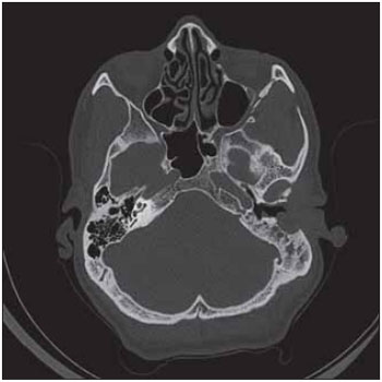
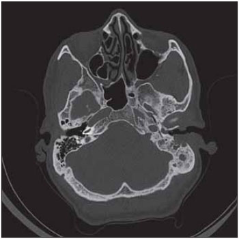
We requested 40-channels mastoid multislice helicoidal computed tomography before and after the venous contrast injection, that revealed a tympanic cavity and antromastoid veiling with destruction of the epitympanum ossicles and wall, temporal bone squamous part and the external auditory meatus, with osseous fragments in the upper region of the external auditory meatus.

Picture 1. CT at axial cut, showing the tympanic cavity veiling and antromastoid with destruction of ossicles.

Picture 2. CT at axial cut, confirming affection of the external auditory meatus.
Then the patient was submitted to left radical mastoidectomy.
The mass removed was forwarded for histopathological exam which revealed a malignant tumor of temporal bone of subtype infiltrative adenoid cystic carcinoma.
After the surgical treatment the patient reported a partial improvement of the facial paralysis, but it culminated in hearing loss because an aggressive procedure was needed for removal of all the mass.
DISCUSSIONOut of the tumors of head and neck, the squamous cells carcinoma is the most common histological type, followed by the basocellular carcinoma, adenoid cystic carcinoma, adenocarcinoma and rhabdomyosarcoma (9).
The adenoid cystic carcinoma is the most common of glandular carcinomas (9). In a review by Conley and Schüller, out of 61 patients with malignant tumors of the external auditory meatus, 19.6% were adenoid cystic carcinoma (1).
The perineural invasion is one of the most treacherous and insidious forms of tumoral dissemination. Because of the extensive neural system, the head and neck malignant tumors have many ways to invade the cranial nerves and gain access to intracranial structures. The most common tumor associated to perineural invasion was the squamous cells carcinoma, followed by the adenoid cystic carcinoma (10). About 30 to 45% of the patients with perineural invasion are initially asymptomatic; the radiologist plays a critical role for subclinical detection of the disease (10).
According to the literature, the symptomatology of this type of tumor is variable and depends basically on the lesion's location. Normally, the patient presents with vague and nonspecific complaints. Head weight, ear pressure, tinnitus, pain, otorrhea or hypacusis may be mentioned. Otalgia is particularly common in the adenoid cystic carcinoma cases for their tendency to invade nerves. Sometimes there are reports of progressive or episodic facial paralysis. In the above mentioned case, the main complaint of the patient was of progressive facial paralysis (4).
Upon otoscopy, in general, we note a grayish or yellowish mass in the external auditory meatus, or, more rarely, medial to the integral tympanic membrane, but the normal otoscopic exam (in cases of tumor out of the meatus) may make the diagnostic suspicion very difficult. In some patients, a narrowing of the meatus is detected (11). Therefore, as presented in the literature, there was no alteration in the otoscopy of the described patient, but the tumor projected onto the mastoid.
Only the histopathological study may state the nature of the tumor with certainty (4).
The surgical treatment of malignant tumors of the ear and temporal bone is not universally standardized. In general, lesions localized in the outer part of the meatus are treated with a limited resection, that is, local resection, radical mastoidectomy, while more advanced lesions are treated by means of block resection, partial temporal osseous resection, subtotal petrosectomy, total petrosectomy. The radiotherapy is defended as a compliment for surgery or a palliative and not as a curative isolated treatment (12). The patient was submitted to radical mastoidectomy and forwarded to the radiotherapy service.
The bad prognosis factors are extensive tumor, facial nerve paralysis, cervical and parotid lymph-node-megaly, as well as invasion of the middle ear, which, when occurs, leads to reduction of the five-year disease-free survival of 59% to 23% (3). In the case in question, the patient presented two factors of bad prognosis: paralysis of the facial nerve and invasion to the middle ear.
FINAL COMMENTSThe adenoid cystic carcinoma is an extremely invasive tumor and, if early diagnosed in its evolution, it presents a better prognosis. In the case report, the tumor was diagnosed at a more advanced phase, partially due to the not much common symptom, peripheral facial paralysis, presented by the patient.
Bibliographical References1. Rapoport PB, Cruz CHG, Lima JB, Carvalho MG. Carcinoma Adenóide Cístico de Conduto Auditivo Externo: Relato de Caso. Rev Bras Otorrinol. 1999, 65(4):1536.
2. Moody SA, Hirsch BE, Myers EN. Squamous Cell Carcinoma of the External Auditory Canal: An Evaluation of a Staging System. The American Journal of Otology. 2000, 21(4):582-588.
3. Gonzalez FM, Paes AJOJ, Tomin OS, Souza RP. Carcinoma Espinocelular do Conduto Auditivo Externo: Estudo por Tomografia Computadorizada de Seis Casos. Rev Bras Radiol. 2005, 38(3):181-185.
4. Caldas SN, Duprat A, Freitas EB, Bento RF, Caldas N. Adenoma ceruminoso do ouvido médio (revisão da literatura e apresentação de um caso). Rev Bras Otorrinol. 1989, 55(4):179-184.
5. Ko YH, Lee MA, Hong YS, Lee KS, Jung C, Kin YS et al. Prognostic factors affecting the clinical outcome of adenoid cystic carcinoma of the head and neck. Jnp J Clin Oncol. 2007, 37(11):805-811.
6. Gormley WB, Sekhar LN, Wright DC, Olding M, Janecka IP, Snyderman CH et al. Management and longterm outcome of adenoid cystic carcinoma with intracranial extension: A neurosurgical perspective. Neurosurgery. 1996, 38:(6)1105-1113.
7. Lalami Y, Vereecker P, Dequanter D, Lothaire P, Awada A. Glands carcinomas, paranasl sinus cancers and melanoma of the head and neck: an update about rare but challenging tumors. Current Opinion in Oncology. 2006, 18:258-265.
8. Prenen H, Kimpe M, Nuyts S. Salivary gland carcinomas: molecular abnormalities as potencial therpeutic targets. Current Opinion in Oncology. 2008, 20:270-274.
9. Paramás AR, Carrasco RG, Britez OA, Yurrita BS. Malignant tumours of the external auditory canal and of the middle ear. Acta Otorrinolaringol Esp. 2004, 55:470-474.
10. Nemzek WR, Hecht S, Gandour-Edwards R, Donald P, McKennan K. Perineural Spread of Head and Neck Tumors: How Accurate Is MR Imaging? AJNR Am J Neuroradiol. 1998, 19:701-706.
11. Perzin KH, Gullane P, Conley J. Adenoid Cystic Carcinoma Involving the External Auditory Canal. Cancer December 15. 1982, 50.
12. Devesa PM, Barnes ML, Milford AC. Malignant Tumors of the Ear and Temporal Bone: A Study of 27 Patients and Review of Their Management. Skull Base. 2008, 18(1).
1. Specialist in Otorhinolaryngology. Coordinator of the Medical Residence Service in Otorhinolaryngology of the Hospital São José do Avaí.
2. Resident of Otorhinolaryngology of the HSJA.
3. Otorhinolaryngology Trainee at HSJA.
Institution: Hospital São Jose do Avaí. Itaperuna / RJ - Brasil.
Mail address:
Paulo Tinoco
Rua Major Porfírio Henriques, 240 - Centro
Itaperuna / RJ - Brasil - Zip code: 28300-000
Telefone: (+55 21) 3822-2836
E-mail: paulo_tinoco@ig.com.br
Article received on July 22 2008.
Accepted on February 09 2009.